
Page 244 - Biodegradable Polyesters
P. 244
222 9 Environment-Friendly Methods for Converting Biodegradable Polyesters
From Table 9.1, another important conclusion could be drawn regarding the
thickness of the studied fibrils. The microfibrils have diameters around 1 μmand
that of nanofibrils is between 50 and 150 nm. The specific surface is also different
for the two types of fibrils. BET analysis of the scaffolds show that the nanofibril-
2 −1
lar material (PET-Nano) is characterized by the highest surface area, 18.8 m g
(Table 9.1). The adsorption–desorption behavior of this sample corresponds to
pores of meso- and micro-size (>10 nm). In contrast to the PET-Nano sample,
the material comprising microfibrils possesses five times less surface area, namely,
2
of 4 m g −1 (Table 9.1, sample PET-Micro 2), and the size of the cavities formed
is in the macro-range (>50 nm). The microfibrillar PGA scaffold, which possesses
larger surface area (Table 9.1) and similar pore size distribution, shows comparable
behavior.
In conclusion, it should be noted that the amount of the organic solvent
in the final nano- or microfibrillar scaffolds depends strongly on the storage
duration at room conditions. It can be reduced drastically to the level of non-
∘
measurable traces applying vacuum drying at elevated temperature (80 C) for
48 h. Regardless of the fact that the content of the organic solvents could be
drastically reduced (Table 9.1), the best solution to the problems created by
the organic solvent residues would be to avoid their use during the scaffold-
manufacturing process as was recently achieved with the preparation of
PLA-based nanofibrillar–nanoporous scaffolds [25, 26].
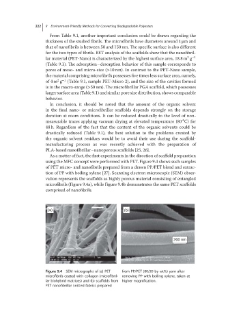
As a matter of fact, the first experiments in the direction of scaffold preparation
using the MFC concept were performed with PET. Figure 9.4 shows such samples
of PET micro- and nanofibrils prepared from a drawn PP/PET blend and extrac-
tion of PP with boiling xylene [27]. Scanning electron microscopic (SEM) obser-
vation represents the scaffolds as highly porous material consisting of entangled
microfibrils (Figure 9.4a), while Figure 9.4b demonstrates the same PET scaffolds
comprised of nanofibrils.
(a) (b)
200 nm
Figure 9.4 SEM micrographs of (a) PET from PP/PET (80/20 by wt%) yarn after
microfibrils coated with collagen (microfibril- removing PP with boiling xylene, taken at
lar biohybrid matrices) and (b) scaffolds from higher magnification.
PET nanofibrillar knitted fabrics prepared