
Page 247 - Biomedical Engineering and Design Handbook Volume 1, Fundamentals
P. 247
224 BIOMECHANICS OF THE HUMAN BODY
Haversian
lamellae Interstitial
Canaliculi lamellae Compact bone
Osteocyte Spongy bone
Lacuna trabeculae
Outer
fibrous layer Haversian
Periosteum canals
Inner
osteogenic layer
Lymphatic vessel in
Haversian canal
Osteoblast Blood vessel in
Volkmann’s canal
Volkmann’s canal
Blood vessels in
Haversian canal
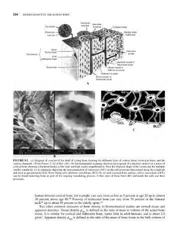
FIGURE 9.2 (a) Diagram of a sector of the shaft of a long bone showing the different types of cortical bone, trabecular bone, and the
various channels. (From Figure 5–1d of Ref. 146.) (b) Environmental scanning electron micrograph of a fracture surface of a piece of
cortical bone showing a fractured lacuna at low (left) and high (right) magnifications. Note the elliptical shape of the lacuna and the multiple
smaller canaliculi. (c) A schematic depicting the interconnection of osteocytes (OC) via the cell processes that extend along the canaliculi
and meet at gap junctions (GJ). Bone-lining cells (dormant osteoblasts, BLC) lie on each exposed bone surface, where osteoclasts (OCL)
can be found removing bone as part of the ongoing remodeling process. A thin layer of bone fluid (BF) surrounds the cells and their
processes.
human femoral cortical bone, for example, can vary from as low as 5 percent at age 20 up to almost
30 percent above age 80. 10 Porosity of trabecular bone can vary from 70 percent in the femoral
11
neck up to about 95 percent in the elderly spine. 12
Two other common measures of bone density in biomechanical studies are termed tissue and
apparent densities. Tissue density r tiss is defined as the ratio of mass to volume of the actual bone
tissue. It is similar for cortical and trabecular bone, varies little in adult humans, and is about 2.0
3
g/cm . Apparent density r is defined as the ratio of the mass of bone tissue to the bulk volume of
app