
Page 248 - Biomedical Engineering and Design Handbook Volume 1, Fundamentals
P. 248
BONE MECHANICS 225
the specimen, including the volume associated with the vascular channels and higher-level porosity.
Volume fraction, tissue density, and apparent densities are related as follows:
r = r V
app tiss f
3
Typically, mean values of apparent density of hydrated cortical bone are about 1.85 g/cm , and
this does not vary much across anatomic sites or species. By contrast, the average apparent density
3
of trabecular bone depends very much on anatomic site. It is as low as 0.10 g/cm for the spine, 13
3
14
3
about 0.30 g/cm for the human tibia, and up to about 0.60 g/cm for the load-bearing portions of
11
the proximal femur. After skeletal maturity (around ages 25 to 30), trabecular bone density decreases
steadily with aging, at a rate of about 6 percent per decade. 15
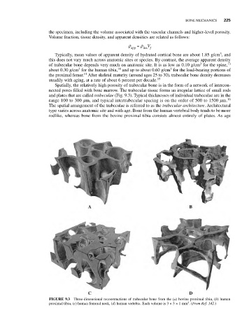
Spatially, the relatively high porosity of trabecular bone is in the form of a network of intercon-
nected pores filled with bone marrow. The trabecular tissue forms an irregular lattice of small rods
and plates that are called trabeculae (Fig. 9.3). Typical thicknesses of individual trabeculae are in the
range 100 to 300 mm, and typical intertrabecular spacing is on the order of 500 to 1500 mm. 16
The spatial arrangement of the trabeculae is referred to as the trabecular architecture. Architectural
type varies across anatomic site and with age. Bone from the human vertebral body tends to be more
rodlike, whereas bone from the bovine proximal tibia consists almost entirely of plates. As age
FIGURE 9.3 Three-dimensional reconstructions of trabecular bone from the (a) bovine proximal tibia, (b) human
3
proximal tibia, (c) human femoral neck, (d) human vertebra. Each volume is 3 × 3 × 1 mm . (From Ref. 142.)