
Page 314 - Biomedical Engineering and Design Handbook Volume 1, Fundamentals
P. 314
ELECTROMYOGRAPHY AS A TOOL TO ESTIMATE MUSCLE FORCES 291
Motor unit force can be explained as a consequence of the cycling of many cross-bridges following
the innervation. Since the stimulus to a motor unit is typically a superposition of several action poten-
tials, a motor unit rarely generates an individual twitch in response to a single action potential. Instead,
these superposed action potentials result in overlapping twitch responses, which is known as tetanus.
When the stimulating frequency at which the action potentials are sent to the motor unit is small, the
tetanus has an irregular force profile. With the increase of the stimulating frequency, the tetanus is
changed to a smooth plateau and has a greater force output. The stimulating frequency can determine
both the magnitude and the shape of the motor unit force profile. Thus, a muscle can increase its force
output by either recruiting more motor units or increasing firing rate.
Electrodes can be used to detect the electrical waveform over a muscle fiber when an action potential
propagates along the muscle fiber, and this waveform is referred to as muscle fiber action potential. Motor
unit action potential (MUAP) is defined as the detected waveform consisting of the spatiotemporal
summation of individual muscle fiber action potentials originating from muscle fibers in the vicinity of a
given electrode or electrode pair. Motor unit action potential train (MUAPT) is defined as the repetitive
sequence of MUAPs from a given motor unit (Winter et al., 1980).
12.2.2 Different Types of Electrodes
The electrodes used to measure EMG are of a wide variety of types and structures. They must be posi-
tioned close enough to the muscle of interest to pick up the electrical signal. Surface electrode, percu-
taneous (needle and wire) electrode are the electrodes usually used (Basmajian and De Luca, 1985).
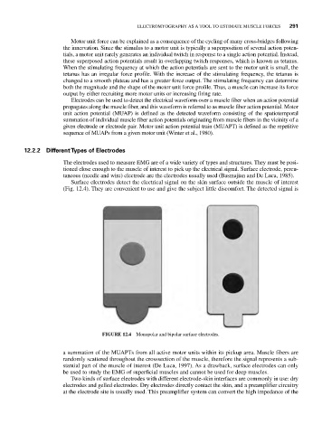
Surface electrodes detect the electrical signal on the skin surface outside the muscle of interest
(Fig. 12.4). They are convenient to use and give the subject little discomfort. The detected signal is
FIGURE 12.4 Monopolar and bipolar surface electrodes.
a summation of the MUAPTs from all active motor units within its pickup area. Muscle fibers are
randomly scattered throughout the crosssection of the muscle, therefore the signal represents a sub-
stantial part of the muscle of interest (De Luca, 1997). As a drawback, surface electrodes can only
be used to study the EMG of superficial muscles and cannot be used for deep muscles.
Two kinds of surface electrodes with different electrode-skin interfaces are commonly in use: dry
electrodes and gelled electrodes. Dry electrodes directly contact the skin, and a preamplifier circuitry
at the electrode site is usually used. This preamplifier system can convert the high impedance of the