
Page 316 - Biomedical Engineering and Design Handbook Volume 1, Fundamentals
P. 316
ELECTROMYOGRAPHY AS A TOOL TO ESTIMATE MUSCLE FORCES 293
The two signals detected at the two different positions are then passed through a differential amplifier
at the electrode site, which treats each signal equally and amplifies the difference between the two
signals. Through this method, the components common in the two signals (mostly noises) are sup-
pressed. Since the electrochemical events in muscle contraction are localized, the electrical signals
emanated from the muscle of interest are different on the two recording positions, and thus can be kept
after passing through the differential amplifier. The noises from a more distant source (the cross-talk
of other muscles, A/C power devices, electromagnetic devices, etc.) are detected with similar ampli-
tudes at both recording positions, and they are treated as correlated signal contents common to both
sites and subtracted prior to being amplified. This way, bipolar electrodes can theoretically eliminate
the noises from distant sources, and they have been used more with the technical development of
electronics amplifiers.
12.2.4 Selection of Different Electrodes
We have introduced many different types and structures of electrodes above. The selection of the
electrodes depends on the practical condition and specific aim.
Surface electrodes are most often used to measure the EMG of superficial large muscles. They are
noninvasive and convenient to use. The data collection can be operated by a person with little training.
Hence surface electrodes are the most popular in a laboratory environment. The bipolar configuration
is recommended to reduce the noises from cross-talk or other distant sources. However, surface elec-
trodes cannot be used for deep muscles, and it is also difficult to use them on small muscles, because
the cross-talk from adjacent muscles would severely contaminate the signal (Robertson, 2004).
Needle and fine wire electrodes must be used to accurately measure the EMG of deep muscles,
small muscles, individual motor units, or muscle fibers. They are invasive and should be used by a
person with special training for inserting these indwelling electrodes. The implementation of needle
and wire electrodes will increase the time and expense of data collection, and may also influence the
subject’s movement pattern because of the pain (especially for needle electrodes).
12.2.5 Electronics of the Electrode System
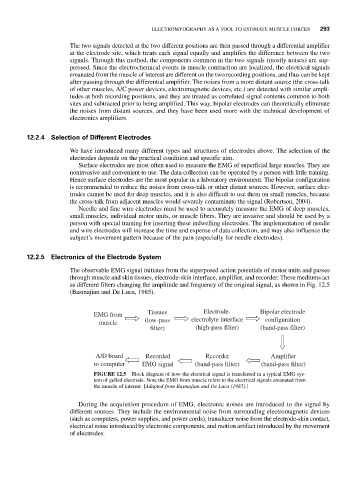
The observable EMG signal initiates from the superposed action potentials of motor units and passes
through muscle and skin tissues, electrode-skin interface, amplifier, and recorder. These mediums act
as different filters changing the amplitude and frequency of the original signal, as shown in Fig. 12.5
(Basmajian and De Luca, 1985).
Tissues Electrode- Bipolar electrode
EMG from
(low-pass electrolyte interface configuration
muscle
filter) (high-pass filter) (band-pass filter)
A/D board Recorded Recorder Amplifier
to computer EMG signal (band-pass filter) (band-pass filter)
FIGURE 12.5 Block diagram of how the electrical signal is transferred in a typical EMG sys-
tem of gelled electrode. Note the EMG from muscle refers to the electrical signals emanated from
the muscle of interest. [Adapted from Basmajian and De Luca (1985).]
During the acquisition procedure of EMG, electronic noises are introduced to the signal by
different sources. They include the environmental noise from surrounding electromagnetic devices
(such as computers, power supplies, and power cords), transducer noise from the electrode-skin contact,
electrical noise introduced by electronic components, and motion artifact introduced by the movement
of electrodes.