Page 254 - Biomedical Engineering and Design Handbook Volume 2, Applications
P. 254
DESIGN OF MAGNETIC RESONANCE SYSTEMS  233

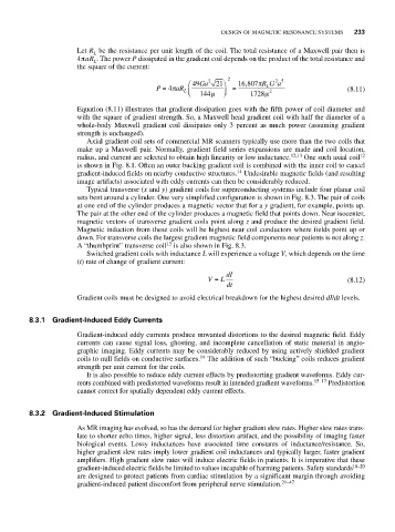
                          Let R be the resistance per unit length of the coil. The total resistance of a Maxwell pair then is
                              L
                          4πaR . The power P dissipated in the gradient coil depends on the product of the total resistance and
                              L
                          the square of the current:
                                                                           25
                                                     ⎛ 49 Ga 2  21 ⎞  2  16 807π R G a
                                                                    ,
                                             P = 4π aR L ⎜    ⎟  =       L                   (8.11)
                                                     ⎝  144μ  ⎠      1728μ  2
                                                                         μ
                          Equation (8.11) illustrates that gradient dissipation goes with the fifth power of coil diameter and
                          with the square of gradient strength. So, a Maxwell head gradient coil with half the diameter of a
                          whole-body Maxwell gradient coil dissipates only 3 percent as much power (assuming gradient
                          strength is unchanged).
                            Axial gradient coil sets of commercial MR scanners typically use more than the two coils that
                          make up a Maxwell pair. Normally, gradient field series expansions are made and coil location,
                          radius, and current are selected to obtain high linearity or low inductance. 12,13  One such axial coil 12
                          is shown in Fig. 8.1. Often an outer bucking gradient coil is combined with the inner coil to cancel
                                                                  14
                          gradient-induced fields on nearby conductive structures. Undesirable magnetic fields (and resulting
                          image artifacts) associated with eddy currents can then be considerably reduced.
                            Typical transverse (x and y) gradient coils for superconducting systems include four planar coil
                          sets bent around a cylinder. One very simplified configuration is shown in Fig. 8.3. The pair of coils
                          at one end of the cylinder produces a magnetic vector that for a y gradient, for example, points up.
                          The pair at the other end of the cylinder produces a magnetic field that points down. Near isocenter,
                          magnetic vectors of transverse gradient coils point along z and produce the desired gradient field.
                          Magnetic induction from these coils will be highest near coil conductors where fields point up or
                          down. For transverse coils the largest gradient magnetic field components near patients is not along z.
                                                13
                          A “thumbprint” transverse coil is also shown in Fig. 8.3.
                            Switched gradient coils with inductance L will experience a voltage V, which depends on the time
                          (t) rate of change of gradient current:
                                                               dI
                                                          V =  L                             (8.12)
                                                               dt
                          Gradient coils must be designed to avoid electrical breakdown for the highest desired dI/dt levels.


              8.3.1 Gradient-Induced Eddy Currents
                          Gradient-induced eddy currents produce unwanted distortions to the desired magnetic field. Eddy
                          currents can cause signal loss, ghosting, and incomplete cancellation of static material in angio-
                          graphic imaging. Eddy currents may be considerably reduced by using actively shielded gradient
                                                        14
                          coils to null fields on conductive surfaces. The addition of such “bucking” coils reduces gradient
                          strength per unit current for the coils.
                            It is also possible to reduce eddy current effects by predistorting gradient waveforms. Eddy cur-
                          rents combined with predistorted waveforms result in intended gradient waveforms. 15–17  Predistortion
                          cannot correct for spatially dependent eddy current effects.


              8.3.2 Gradient-Induced Stimulation
                          As MR imaging has evolved, so has the demand for higher gradient slew rates. Higher slew rates trans-
                          late to shorter echo times, higher signal, less distortion artifact, and the possibility of imaging faster
                          biological events. Lossy inductances have associated time constants of inductance/resistance. So,
                          higher gradient slew rates imply lower gradient coil inductances and typically larger, faster gradient
                          amplifiers. High gradient slew rates will induce electric fields in patients. It is imperative that these
                          gradient-induced electric fields be limited to values incapable of harming patients. Safety standards 18–20
                          are designed to protect patients from cardiac stimulation by a significant margin through avoiding
                          gradient-induced patient discomfort from peripheral nerve stimulation. 21–47

