
Page 382 - Biomedical Engineering and Design Handbook Volume 2, Applications
P. 382
360 DIAGNOSTIC EQUIPMENT DESIGN
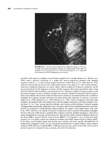
FIGURE 12.5 Post Gd contrast enhanced Ts subtraction image in a 40-year-
old high-risk female demonstrates nonmass-like enhancement in the upper out
quadrant of the left breast that was confirmed histologically to be segmental
involvement with DCIS. Mammogram was normal.
and differ with respect to emphasis on information acquired. For example, Boetes et al. (Boetes et al.,
1994) used a protocol consisting of a single-slice non-fat-suppressed gradient echo imaging
sequence with 2.6- × 1.3-mm in-plane spatial resolution (10-mm slice) at 2.3-s time intervals. They
used the criterion that any lesion with visible enhancement in less than 11.5 s after arterial enhance-
ment was considered suspicious for cancer. These criteria resulted in 95 percent sensitivity and 86
percent specificity for the diagnosis of cancer. Similar sequences have been reported by others using
protocols with the time resolution varying from 6 to 60 s. However, problems locating a lesion on the
precontrast images in order to perform a single-slice dynamic enhanced examination and the need to
detect and evaluate other lesions within the breast have resulted in recommendations that a multislice
technique that captures dynamic data from the entire breast after injection of contrast is necessary
(Gilles et al., 1994; Hickman et al., 1994; Perman et al., 1994). These investigators advocate using
multislice 2D gradient echo, 3D gradient echo, and echo planar techniques, with time resolution vary-
ing from 12 s to 1 min, varying special resolution, and varying section thickness. Keyhole imaging
techniques that dynamically sample the center of k-space after contrast administration have been sug-
gested as a technique to obtain dynamic high-resolution 3D images of the entire breast (Van Vaals
et al., 1993). However, the spatial resolution of enhanced tissue is limited with keyhole techniques
because only part of the breast is sampled after contrast is injected. Keyhole imaging is criticized as
being suboptimal for assessing lesion architecture. Research to clarify optimal acquisition protocols
for breast MRI is needed. Recent work in breast MRI in 3-T magnets is very exciting and holds
promise for even higher spatial and temporal resolution and further improvements in image quality.
In the future, novel contrast agents may provide more sensitive and more specific discrimination of
benign from malignant lesions. In vivo functional measurements of tumor biology using contrast-
enhanced MRI, diffusion-weighted MRI, or MR spectroscopy may yield markers that can be used to
predict response to treatment more accurately and earlier in treatment.