
Page 377 - Biomedical Engineering and Design Handbook Volume 2, Applications
P. 377
BREAST IMAGING SYSTEMS: DESIGN CHALLENGES FOR ENGINEERS 355
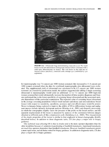
FIGURE 12.3 Ultrasound image demonstrating a mass and a cyst. The round
lesion on the left demonstrates homogenous internal echoes consistent with a
solid mass (fibroadenoma on biopsy). The oval lesion on the right has no
internal echoes (anechoic), consistent with a benign cyst (confirmed by cyst
aspiration).
for mammography was 7.6 cancers per 1000 women screened; this increased to 11.8 cancers per
1000 women screened when the data for combined mammography plus ultrasound were evalu-
ated. The supplemental yield of ultrasound was calculated to be 4.2 cancers per 1000 women
screened. In a sensitivity predication model, the authors suggested that adding a single screening
ultrasound to mammography would yield an additional 1.1 to 7.2 cancers per 1000 high-risk
women screened but would substantially increase the number of false positive diagnoses and biop-
sies performed due to the lower specificity of screening breast ultrasound and the associated oper-
ator dependence of this particular examination. The expected value of screening breast ultrasound
in the average screening population (which would include radiodense and non-radiodense breast
tissue) with respect to sensitivity, specificity, accuracy and cost-effectiveness would be poorer.
Newer techniques being explored to improve the ability of US to differentiate benign and malig-
nant masses include intensity histogram analysis (Kitaoka et al., 2001) and disparity processing,
in which the sonographer slightly varies the pressure of the probe on the breast surface, and the
apparent displacement of the tissue is measured by analysis of the correlation between images
obtained at different parts of this compression cycle (Steinberg et al., 2001). This measurement
of the elastic properties of the lesion is similar to that employed in breast elastography (briefly
described below). In addition to these diagnostic tasks, US also plays a major role in biopsy
guidance.
One technical issue affecting US is that its results tend to be more operator-dependent than the
other modalities because of variations in positioning of handheld transducers. Automated transducers
are much less operator-dependent than handheld transducers. However, handheld transducers permit
a more rapid exam, and are better suited for biopsy guidance. In addition to diagnostic tasks, US also
plays a major role in biopsy guidance.