
Page 372 - Biomedical Engineering and Design Handbook Volume 2, Applications
P. 372
350 DIAGNOSTIC EQUIPMENT DESIGN
successive imaging studies is difficult, if not impossible. This complicates correlation between fol-
low-up images of a given modality, or between concurrently obtained images from two different
modalities. A second challenge arises from the fact that cancers can arise in areas of the breast very
close to the chest wall. For example, the focal spot in x-ray mammography must be positioned
directly above the chest wall edge of the image receptor in order to assure that x-rays passing through
tissue adjacent to the chest wall are imaged. The proximity of the chest and shoulders also presents
geometric hindrance affecting MRI coil design and nuclear medicine scanning. A third challenging
aspect of breast cancer imaging is the similarity of many of the physical attributes of cancerous mate-
rial and normal breast tissue. For example, the x-ray attenuation and acoustic impedance of cancer-
ous masses are very similar to those of healthy fibroglandular breast tissue. Thus the imaging process
must result in a sufficiently high signal-to-noise ratio that such subtle differences can be ascertained.
12.2 BREAST ANATOMY
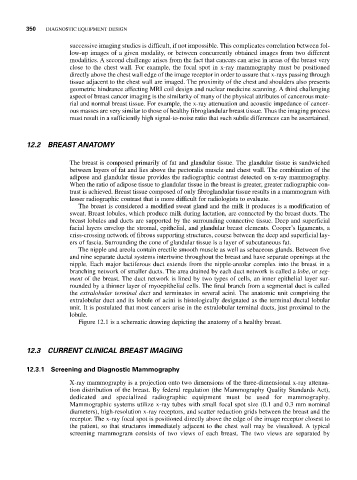
The breast is composed primarily of fat and glandular tissue. The glandular tissue is sandwiched
between layers of fat and lies above the pectoralis muscle and chest wall. The combination of the
adipose and glandular tissue provides the radiographic contrast detected on x-ray mammography.
When the ratio of adipose tissue to glandular tissue in the breast is greater, greater radiographic con-
trast is achieved. Breast tissue composed of only fibroglandular tissue results in a mammogram with
lesser radiographic contrast that is more difficult for radiologists to evaluate.
The breast is considered a modified sweat gland and the milk it produces is a modification of
sweat. Breast lobules, which produce milk during lactation, are connected by the breast ducts. The
breast lobules and ducts are supported by the surrounding connective tissue. Deep and superficial
facial layers envelop the stromal, epithelial, and glandular breast elements. Cooper’s ligaments, a
criss-crossing network of fibrous supporting structures, course between the deep and superficial lay-
ers of fascia. Surrounding the cone of glandular tissue is a layer of subcutaneous fat.
The nipple and areola contain erectile smooth muscle as well as sebaceous glands. Between five
and nine separate ductal systems intertwine throughout the breast and have separate openings at the
nipple. Each major lactiferous duct extends from the nipple-areolar complex into the breast in a
branching network of smaller ducts. The area drained by each duct network is called a lobe, or seg-
ment of the breast. The duct network is lined by two types of cells, an inner epithelial layer sur-
rounded by a thinner layer of myoepithelial cells. The final branch from a segmental duct is called
the extralobular terminal duct and terminates in several acini. The anatomic unit comprising the
extralobular duct and its lobule of acini is histologically designated as the terminal ductal lobular
unit. It is postulated that most cancers arise in the extralobular terminal ducts, just proximal to the
lobule.
Figure 12.1 is a schematic drawing depicting the anatomy of a healthy breast.
12.3 CURRENT CLINICAL BREAST IMAGING
12.3.1 Screening and Diagnostic Mammography
X-ray mammography is a projection onto two dimensions of the three-dimensional x-ray attenua-
tion distribution of the breast. By federal regulation (the Mammography Quality Standards Act),
dedicated and specialized radiographic equipment must be used for mammography.
Mammographic systems utilize x-ray tubes with small focal spot size (0.1 and 0.3 mm nominal
diameters), high-resolution x-ray receptors, and scatter reduction grids between the breast and the
receptor. The x-ray focal spot is positioned directly above the edge of the image receptor closest to
the patient, so that structures immediately adjacent to the chest wall may be visualized. A typical
screening mammogram consists of two views of each breast. The two views are separated by