
Page 367 - Biomedical Engineering and Design Handbook Volume 2, Applications
P. 367
NUCLEAR MEDICINE IMAGING INSTRUMENTATION 345
A B
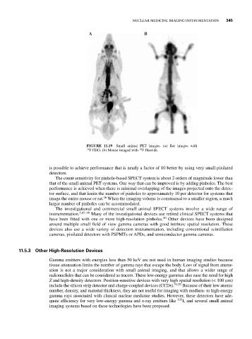
FIGURE 11.19 Small animal PET images. (a) Rat images with
18 F FDG. (b) Mouse imaged with F fluoride.
18
is possible to achieve performance that is nearly a factor of 10 better by using very small pixilated
detectors.
The count sensitivity for pinhole-based SPECT system is about 2 orders of magnitude lower than
that of the small animal PET systems. One way that can be improved is by adding pinholes. The best
performance is achieved when there is minimal overlapping of the images projected onto the detec-
tor surface, and that limits the number of pinholes to approximately 10 per detector for systems that
46
image the entire mouse or rat. When the imaging volume is constrained to a smaller region, a much
larger number of pinholes can be accommodated.
The investigational and commercial small animal SPECT systems involve a wide range of
instrumentation. 5,47–49 Many of the investigational devices use retired clinical SPECT systems that
have been fitted with one or more high-resolution pinholes. 50 Other devices have been designed
around multiple small field of view gamma cameras with good intrinsic spatial resolution. These
devices also use a wide variety of detection instrumentation, including conventional scintillation
cameras, pixilated detectors with PSPMTs or APDs, and semiconductor gamma cameras.
11.5.3 Other High-Resolution Devices
Gamma emitters with energies less than 50 keV are not used in human imaging studies because
tissue attenuation limits the number of gamma rays that escape the body. Loss of signal from attenu-
ation is not a major consideration with small animal imaging, and that allows a wider range of
radionuclides that can be considered as tracers. These low-energy gammas also ease the need for high
Z and high-density detectors. Position-sensitive devices with very high spatial resolution (< 100 μm)
include the silicon strip detector and charge-coupled devices (CCDs). 51,52 Because of their low atomic
number, density, and material thickness, they are not useful for imaging with medium- to high-energy
gamma rays associated with clinical nuclear medicine studies. However, these detectors have ade-
quate efficiency for very low-energy gamma and x-ray emitters like 125 I, and several small animal
imaging systems based on these technologies have been proposed.