
Page 364 - Biomedical Engineering and Design Handbook Volume 2, Applications
P. 364
342 DIAGNOSTIC EQUIPMENT DESIGN
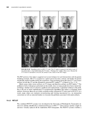
FIGURE 11.18 Breathing artifact on PET CT study. The CT study is acquired in the helical mode. If
the patient takes a deep breath during the CT study, a “floating liver” artifact often occurs. Because the
CT is used for attenuation correction, the artifact is also evident on the PET images.
The PET portion of the study is acquired over several minutes for each bed position with the patient
breathing normally. The CT is acquired as a spiral scan in less than a minute, and ideally the patient
should take shallow breaths during the acquisition. Deep breathing during the spiral CT scan creates
artifacts in both the CT and the attenuation corrected PET studies 32,33 as shown in Fig. 11.18.
34
Metal causes rather severe artifacts on CT images and therefore artifacts on PET studies. The
problem is fairly widespread, particularly in the mouth, since so many people have heavy metal den-
tal fillings. Another area of concern is artificial joint replacements. A potential solution to this prob-
lem is the use of more sophisticated CT reconstruction algorithms that reduce or eliminate metal
artifacts. However, these are not widely available on the commercial CT units used in PET/CT. As a
result, many clinics are routinely reconstructing and viewing uncorrected (i.e., no attenuation
correction) as well as corrected studies to overcome the problems associated with metal artifacts.
11.4.2 PET/CT
The combined PET/CT systems were developed at the University of Pittsburgh by Townsend et al.
and were initially introduced as commercial devices in 2001. 35–37 These devices quickly caught on
and have virtually replaced all the stand-alone PET tomographs. The PET/CT systems combine a