
Page 359 - Biomedical Engineering and Design Handbook Volume 2, Applications
P. 359
NUCLEAR MEDICINE IMAGING INSTRUMENTATION 337
A B
A B PMTs
Detectors
Light
C D pipe
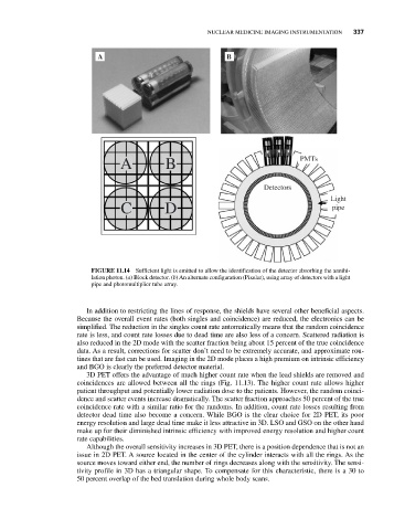
FIGURE 11.14 Sufficient light is emitted to allow the identification of the detector absorbing the annihi-
lation photon. (a) Block detector. (b) An alternate configuration (Pixelar), using array of detectors with a light
pipe and photomultiplier tube array.
In addition to restricting the lines of response, the shields have several other beneficial aspects.
Because the overall event rates (both singles and coincidence) are reduced, the electronics can be
simplified. The reduction in the singles count rate automatically means that the random coincidence
rate is less, and count rate losses due to dead time are also less of a concern. Scattered radiation is
also reduced in the 2D mode with the scatter fraction being about 15 percent of the true coincidence
data. As a result, corrections for scatter don’t need to be extremely accurate, and approximate rou-
tines that are fast can be used. Imaging in the 2D mode places a high premium on intrinsic efficiency
and BGO is clearly the preferred detector material.
3D PET offers the advantage of much higher count rate when the lead shields are removed and
coincidences are allowed between all the rings (Fig. 11.13). The higher count rate allows higher
patient throughput and potentially lower radiation dose to the patients. However, the random coinci-
dence and scatter events increase dramatically. The scatter fraction approaches 50 percent of the true
coincidence rate with a similar ratio for the randoms. In addition, count rate losses resulting from
detector dead time also become a concern. While BGO is the clear choice for 2D PET, its poor
energy resolution and large dead time make it less attractive in 3D. LSO and GSO on the other hand
make up for their diminished intrinsic efficiency with improved energy resolution and higher count
rate capabilities.
Although the overall sensitivity increases in 3D PET, there is a position dependence that is not an
issue in 2D PET. A source located in the center of the cylinder interacts with all the rings. As the
source moves toward either end, the number of rings decreases along with the sensitivity. The sensi-
tivity profile in 3D has a triangular shape. To compensate for this characteristic, there is a 30 to
50 percent overlap of the bed translation during whole body scans.