
Page 360 - Biomedical Engineering and Design Handbook Volume 2, Applications
P. 360
338 DIAGNOSTIC EQUIPMENT DESIGN
Fair comparisons of 2D and 3D PET modes cannot be made solely on the basis of the measured
count rates, since the increase in count rate in 3D PET is accompanied by increases in the magnitude
of corrections for both randoms and scatter. To effect meaningful comparisons, the concept of the
noise equivalent count rate (NECR) is used. The noise equivalent count rate is a way of comparing
the actual gain associated with an increased sensitivity that also requires increased corrections. The
NECR is expressed in terms of the true coincidence count rate (the good data, usually represented
by T), the scatter count rate (S), and the random coincidence rate (R):
2
NECR = T /(T + S + cR)
where c is a constant that is equal to either 1 or 2 depending on how the random coincidence rate is
determined.
For 2D PET, the scatter fraction is about 10 percent of the acquired events and a typical random
coincidence rate is 25 percent of the acquired events. In 3D PET both the scatter fraction and the ran-
dom coincidence rate are close to 50 percent of the true coincidence rate. So even though the
observed rate goes up by more than a factor of 6 in going from 2D to 3D PET, the improvement in
NEC is between 3 and 4. The NEC is also useful in comparing the count rate performance of the dif-
ferent 3D PET systems.
One of the advantages of PET imaging is its relatively good spatial resolution compared with
SPECT. The primary factors that influence spatial resolution include the face size of the detectors, the
detector thickness, the detector separation, data smoothing during or after reconstruction, and the
27
pixel size of the displayed images. PET tomographs achieve spatial resolution through coincidence
detection. Since the two annihilation photons must strike the opposed coincidence detectors, the spa-
tial resolution associated with the LOR is equal to the half detector face size. This, of course, depends
on being able to accurately identify individual detector elements. Because of the uncertainty involved
in selecting the correct detector with each event, the spatial resolution is approximately equal to the
detector face size.
To achieve high intrinsic efficiency, the detectors used in PET tomographs are typically 20 to
30 mm thick. When an annihilation photon strikes the detector, it can be absorbed anywhere along
the detector length. Annihilation photons arriving from the center of the tomograph field of view are
likely to travel along the axis of a detector and this presents no problem. However, annihilation pho-
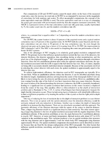
tons coming from the edge of the field of view have trajectories that cross many detectors. Because
the exact location of the interaction along the detector is unknown, the LOR is essentially broadened,
leading to a significant loss of spatial resolution that increases as the source location is displaced
from the center of the ring. This parallax effect is often referred to as the depth of interaction
problem and is illustrated in Fig. 11.15. A variety of techniques have been proposed to measure the
depth that the interaction occurs. These include using layers of detectors with identifiable differences
in their scintillation properties or by treating the surface of the crystals so that the light yield is depth
dependent. 29
One way of reducing the loss of resolution due to this depth of interaction problem is to use thinner
detectors. This is not an option with the current detectors since the detection efficiency would be
seriously reduced with any reduction in thickness. Another way to reduce the depth of interaction
problem is to increase the ring diameter. There are two drawbacks associated with that approach. The
geometric efficiency of the detectors decreases with the square of the source to detector distance.
Thus, a larger ring diameter results in a significant drop in count sensitivity. The other reason oppos-
ing a larger ring diameter is the loss of spatial resolution associated with the angular dispersion of the
annihilation photons. If the positron annihilates at rest, the angle between the 2 annihilation photons
o
o
is exactly 180 . However, at room (and body) temperature, thermal motion adds about 0.5 (± 0.25)
variation in the annihilation angle. This variation produces a loss in spatial resolution that increases
with the ring diameter and is approximately 2 mm for whole body PET tomographs (ring diameter
approximately 90 cm). The spatial resolution components discussed above can be described as intrin-
sic because they are fixed by the design configuration of the PET tomograph. The other factors that
influence spatial resolution, smoothing, and pixel size vary, depending on reconstruction parameter
selections.