
Page 361 - Biomedical Engineering and Design Handbook Volume 2, Applications
P. 361
NUCLEAR MEDICINE IMAGING INSTRUMENTATION 339
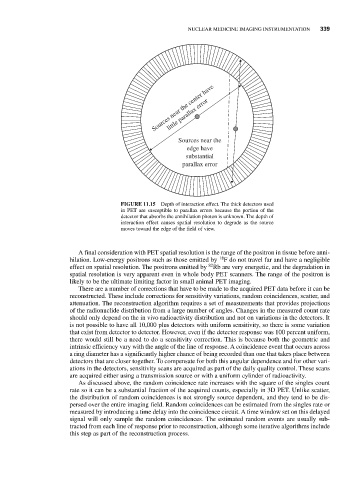
Sources near the center have
little parallax error
Sources near the
edge have
substantial
parallax error
FIGURE 11.15 Depth of interaction effect. The thick detectors used
in PET are susceptible to parallax errors because the portion of the
detector that absorbs the annihilation photon is unknown. The depth of
interaction effect causes spatial resolution to degrade as the source
moves toward the edge of the field of view.
A final consideration with PET spatial resolution is the range of the positron in tissue before anni-
18
hilation. Low-energy positrons such as those emitted by F do not travel far and have a negligible
82
effect on spatial resolution. The positrons emitted by Rb are very energetic, and the degradation in
spatial resolution is very apparent even in whole body PET scanners. The range of the positron is
likely to be the ultimate limiting factor in small animal PET imaging.
There are a number of corrections that have to be made to the acquired PET data before it can be
reconstructed. These include corrections for sensitivity variations, random coincidences, scatter, and
attenuation. The reconstruction algorithm requires a set of measurements that provides projections
of the radionuclide distribution from a large number of angles. Changes in the measured count rate
should only depend on the in vivo radioactivity distribution and not on variations in the detectors. It
is not possible to have all 10,000 plus detectors with uniform sensitivity, so there is some variation
that exist from detector to detector. However, even if the detector response was 100 percent uniform,
there would still be a need to do a sensitivity correction. This is because both the geometric and
intrinsic efficiency vary with the angle of the line of response. A coincidence event that occurs across
a ring diameter has a significantly higher chance of being recorded than one that takes place between
detectors that are closer together. To compensate for both this angular dependence and for other vari-
ations in the detectors, sensitivity scans are acquired as part of the daily quality control. These scans
are acquired either using a transmission source or with a uniform cylinder of radioactivity.
As discussed above, the random coincidence rate increases with the square of the singles count
rate so it can be a substantial fraction of the acquired counts, especially in 3D PET. Unlike scatter,
the distribution of random coincidences is not strongly source dependent, and they tend to be dis-
persed over the entire imaging field. Random coincidences can be estimated from the singles rate or
measured by introducing a time delay into the coincidence circuit. A time window set on this delayed
signal will only sample the random coincidences. The estimated random events are usually sub-
tracted from each line of response prior to reconstruction, although some iterative algorithms include
this step as part of the reconstruction process.