
Page 363 - Biomedical Engineering and Design Handbook Volume 2, Applications
P. 363
NUCLEAR MEDICINE IMAGING INSTRUMENTATION 341
attenuation, while those that traverse the thickest portion of the patient can have path lengths of
greater than 50 cm in large patients, leading to reductions in the coincidence detection rate of more
than a factor of 100. This causes a nonlinear relationship between the detected counts and the source
activity. Thus, reconstruction of the acquired data without attenuation correction leads to the char-
acteristic artifacts, including enhanced count density at the skin and in the lung fields and decreased
count density in the central portions of the patient (Fig. 11.16b).
The amount of attenuation depends only on the trajectory of the annihilation photons through the
patient, not on the actual location of the source. Measurement of the transmission factors can be per-
formed using radionuclide sources that revolve about the patient using the PET tomograph as a crude
68
CT scanner. 11,30 Radionuclide sources that have been used for that application include Ge and
137 Cs. However, collecting the transmission information this way is slow (1 to 3 minutes per bed
position) and produces low-resolution, noisy corrections. These problems have been largely solved
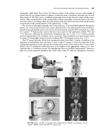
by using CT tomographic images from the combined PET CT imaging systems (Fig. 11.17).
The CT scanner provides whole body transmission data in less than 1 minute. In addition to the
reduction in transmission time, the level of noise in the CT images is much less than that of the
radionuclide transmission data. Because the average energy of the x-rays used in the CT scan is 60 to
80 keV, the CT attenuation coefficients have to be mapped to the appropriate values at 511 keV.
Although this is a nonlinear process, the mapping has been successfully implemented. 31 However,
there are several potential problems that occur when using CT data for attenuation correction.
A B
FIGURE 11.17 (a) PET CT scanners from General Electric, Philips, and Siemens. (b) An
18
example F FDG whole body PET CT clinical study.