
Page 368 - Biomedical Engineering and Design Handbook Volume 2, Applications
P. 368
346 DIAGNOSTIC EQUIPMENT DESIGN
99m Tc bone scan
CT
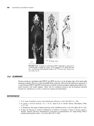
FIGURE 11.20 Example of a small animal SPECT study with co-registered CT.
(a) Whole body CT image of a mouse. (b) Co-registered 99m Tc whole body bone
scan. (c) Maximum pixel projection image of the 99m Tc bone scan. Images cour-
tesy of Bioscan, Inc.
11.6 SUMMARY
Nuclear medicine, including both SPECT and PET, has been on the leading edge of the molecular
imaging revolution. Because of the incredibly high sensitivity offered by the radiotracer approach, it
is expected that SPECT and PET will remain as valuable clinical modalities and irreplaceable for tar-
geted research with small animals. There will be continued research and development directed
toward new radiotracers as well as improved imaging instrumentation.
REFERENCES
1. H. O. Anger, Scintillation camera with multichannel collimators, J Nucl Med, 5:515–31, 1964.
2. D. Gunter, in Nuclear Medicine; Vol. 1, 2d ed., edited by R. E. Henkin (Mosby, Philadelphia, 2006),
p. 107–126.
3. G. Muehllehner, The impact of digital technology on the scintillation camera, J Nucl Med, 22(4):389–91, 1981.
4. H. Hines, R. Kayayan, J. Colsher, D. Hashimoto, R. Schubert, J. Fernando, V. Simcic, P. Vernon, and R. L.
Sinclair, National Electrical Manufacturers Association recommendations for implementing SPECT
instrumentation quality control, J Nucl Med, 41(2):383–9, 2000.