
Page 373 - Biomedical Engineering and Design Handbook Volume 2, Applications
P. 373
BREAST IMAGING SYSTEMS: DESIGN CHALLENGES FOR ENGINEERS 351
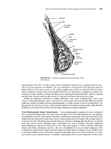
FIGURE 12.1 Schematic diagram showing the anatomy of the
human breast.
approximately 45 to 60° in order to help resolve ambiguities produced by overlapping breast struc-
ture in a given projection. In addition, one view maximizes visualization of the structures near the
lateral portion of the breast, such as the axillary lymph nodes. Both are obtained with the breast
under compression using a flat acrylic paddle with low x-ray attenuation. Compression reduces the
amount of scatter radiation reaching the detector by reducing the breast thickness, and also spreads
out the tissue, thereby reducing the amount of structural overlap in the projection image.
Follow-up diagnostic procedures include additional x-ray views (magnification views, spot
views, or unusual projections such as lateral views), ultrasound, and more recently MRI and nuclear
medicine imaging (positron emission mammography or single gamma emission scintigraphy). We
first present an overview of some of the technical aspects of screening mammography, then describe
challenges associated with current and upcoming diagnostic imaging techniques.
X-ray Mammography Image Considerations. X-ray mammography is perhaps the most exacting of
all x-ray–based imaging tasks. The main reasons for this are (1) the small difference in x-ray attenua-
tion properties between various breast structures, and between normal and cancerous tissue and (2) the
requirement that physically small objects such as microcalcifications be imaged with enough clarity to
be detected by the radiologist (microcalcifications are calcium-containing deposits that are associated
with early breast cancers, although many calcifications are merely benign). Clinically significant
microcalcifications may be 0.2 mm or less in size. They often appear in clusters, and their individual
shapes and relative orientations can be a clue as to the likelihood of an associated malignancy. The
simultaneous requirements of high contrast resolution and high spatial resolution, along with the desire
to minimize radiation dose to the breast, dictate that the image sensor have high sensitivity, low noise,
and a narrow point response function. In addition, depending on the size and composition of the breast,