
Page 375 - Biomedical Engineering and Design Handbook Volume 2, Applications
P. 375
BREAST IMAGING SYSTEMS: DESIGN CHALLENGES FOR ENGINEERS 353
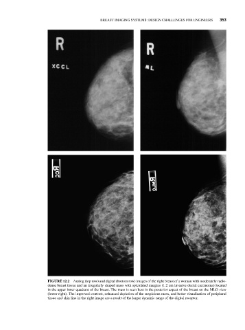
FIGURE 12.2 Analog (top row) and digital (bottom row) images of the right breast of a woman with moderately radio-
dense breast tissue and an irregularly shaped mass with spiculated margins (1.2 cm invasive ductal carcinoma) located
in the upper inner quadrant of the breast. The mass is seen best in the posterior aspect of the breast on the MLO view
(lower right). The improved contrast, enhanced depiction of the suspicious mass, and better visualization of peripheral
tissue and skin line in the right image are a result of the larger dynamic range of the digital receptor.