
Page 354 - Biomedical Engineering and Design Handbook Volume 2, Applications
P. 354
332 DIAGNOSTIC EQUIPMENT DESIGN
11.3.5 Compton Cameras
Collimators are very inefficient and are the limiting factor in conventional scintillation cameras. One
device that eliminates the need for lead collimation is the Compton camera which was investigated
during the 1980s and has resurfaced in the past 5 years. 21–23 Instead of restricting the gamma ray
trajectories, the Compton camera uses scattering information from two position-sensitive detectors
to infer the source location. The gamma ray is Compton scattered from the first detector, and the
scattered photon is totally absorbed in the second detector. The energy of the scattered photon is also
determined by the second detector and that information allows the calculation of the scattering angle
between the incoming gamma ray and the known path between the two detectors. The information
from a single event restricts the source to the surface of a cone, and reconstruction algorithms can
provide tomographic images of the source distributions. Compton cameras are estimated to improve
count sensitivity by a factor of 100; however, useful devices have not yet been developed for clini-
cal imaging. Compton cameras appear to work best with isolated point source distributions and are
challenged with the three-dimensional distribution volumes associated with most nuclear medicine
studies. As a result, the best application for this approach may be small animal imaging. Preclinical
imaging systems designed for small animals utilizing SPECT and PET are reviewed in Sec. 11.5.
11.4 POSITRON EMISSION TOMOGRAPHY
Positron emission tomography (PET) is another approach to nuclear medicine imaging that has sev-
eral advantages over SPECT. As noted in the introduction to this chapter, PET uses positron-emitting
radionuclides that result in the emission of collinear pairs of 511-keV annihilation photons. The coin-
cidence detection of the annihilation photons obviates the need for collimation and makes PET far
more efficient than SPECT for detecting radioactivity. Even more importantly, there are positron-
emitting radionuclides for oxygen, carbon, nitrogen, and fluorine (Table 11.4), which allows a wide
range of molecules to be labeled as diagnostic agents. Many of these radionuclides have short half-
18
lives and require an on-site cyclotron. However, F has a sufficiently long half-life that it can be (and is)
regionally provided, and there is no populated area of the United States where it is unavailable.
68
82
Several others such as Rb and Ga are available from radionuclide generators that provide the
radionuclides on demand despite their short half-lives.
Coincidence detection provides spatial resolution without the need for lead collimation by taking
advantage of the fact that the annihilation photons resulting from positron emission are approximately
colinear. Events are only counted if they are simultaneously detected by two opposed detectors. The
sensitive volume defined by the coincidence detectors is called a line of response (LOR). As illustrated
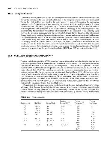
TABLE 11.4 PET Radionuclides
Positron Photon Photons
Radionuclide Half-Life Decay Mode Energy (MeV) Energy (keV) per Decay
11 C 20.4 m β+ 0.96 511 2
13 N 10.0 m β+ 1.19 511 2
15 O 2.0 m β+ 1.72 511 2
18 F 109.8 m β+, EC 0.63 511 1.93
82 Rb 76 s β+, EC 3.35 511 1.9
776 0.13
68 Ga 68.3 m β+, EC 1.9 511 1.84
64 Cu 12.7 h β–, β+, EC 0.65 511 0.38
1346 0.005
124 I 4.2 d β+, EC 1.54, 2.17 511 0.5
603 0.62
1693 0.3