
Page 353 - Biomedical Engineering and Design Handbook Volume 2, Applications
P. 353
NUCLEAR MEDICINE IMAGING INSTRUMENTATION 331
A B
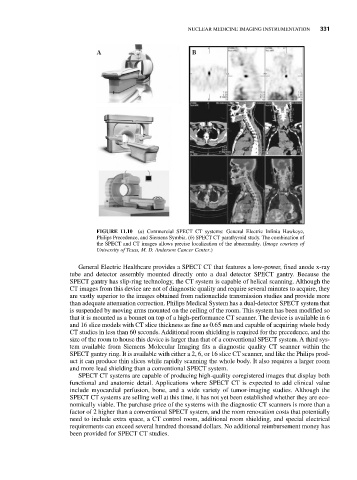
FIGURE 11.10 (a) Commercial SPECT CT systems: General Electric Infinia Hawkeye,
Philips Precedence, and Siemens Symbia. (b) SPECT CT parathyroid study. The combination of
the SPECT and CT images allows precise localization of the abnormality. (Image courtesy of
University of Texas, M. D. Anderson Cancer Center.)
General Electric Healthcare provides a SPECT CT that features a low-power, fixed anode x-ray
tube and detector assembly mounted directly onto a dual detector SPECT gantry. Because the
SPECT gantry has slip-ring technology, the CT system is capable of helical scanning. Although the
CT images from this device are not of diagnostic quality and require several minutes to acquire, they
are vastly superior to the images obtained from radionuclide transmission studies and provide more
than adequate attenuation correction. Philips Medical System has a dual-detector SPECT system that
is suspended by moving arms mounted on the ceiling of the room. This system has been modified so
that it is mounted as a bonnet on top of a high-performance CT scanner. The device is available in 6
and 16 slice models with CT slice thickness as fine as 0.65 mm and capable of acquiring whole body
CT studies in less than 60 seconds. Additional room shielding is required for the precedence, and the
size of the room to house this device is larger than that of a conventional SPECT system. A third sys-
tem available from Siemens Molecular Imaging fits a diagnostic quality CT scanner within the
SPECT gantry ring. It is available with either a 2, 6, or 16 slice CT scanner, and like the Philips prod-
uct it can produce thin slices while rapidly scanning the whole body. It also requires a larger room
and more lead shielding than a conventional SPECT system.
SPECT CT systems are capable of producing high-quality coregistered images that display both
functional and anatomic detail. Applications where SPECT CT is expected to add clinical value
include myocardial perfusion, bone, and a wide variety of tumor-imaging studies. Although the
SPECT CT systems are selling well at this time, it has not yet been established whether they are eco-
nomically viable. The purchase price of the systems with the diagnostic CT scanners is more than a
factor of 2 higher than a conventional SPECT system, and the room renovation costs that potentially
need to include extra space, a CT control room, additional room shielding, and special electrical
requirements can exceed several hundred thousand dollars. No additional reimbursement money has
been provided for SPECT CT studies.