
Page 348 - Biomedical Engineering and Design Handbook Volume 2, Applications
P. 348
326 DIAGNOSTIC EQUIPMENT DESIGN
11.3 SINGLE PHOTON EMISSION COMPUTED TOMOGRAPHY
SPECT (single photon emission computed tomography) produces tomographic images of the
10
internal distribution of radiopharmaceuticals. It is most commonly used in the diagnosis of coro-
nary artery disease and in tumor detection. Projection images collected by one or more scintilla-
tion cameras are mathematically reconstructed to obtain the tomographic slices. SPECT studies
are performed for a wide variety of clinical indications and are often used in the diagnosing and
monitoring of malignancies. However, myocardial perfusion studies evaluating the heart for coro-
nary artery disease are by far the most common SPECT procedures. Quantitative SPECT yielding
absolute radioactivity concentrations requires corrections for attenuation, scatter, and spatial
resolution.
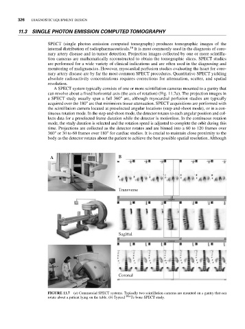
A SPECT system typically consists of one or more scintillation cameras mounted to a gantry that
can revolve about a fixed horizontal axis (the axis of rotation) (Fig. 11.7a). The projection images in
a SPECT study usually span a full 360° arc, although myocardial perfusion studies are typically
acquired over the 180° arc that minimizes tissue attenuation. SPECT acquisitions are performed with
the scintillation camera located at preselected angular locations (step-and-shoot mode), or in a con-
tinuous rotation mode. In the step-and-shoot mode, the detector rotates to each angular position and col-
lects data for a preselected frame duration while the detector is motionless. In the continuous rotation
mode, the study duration is selected and the rotation speed is adjusted to complete the orbit during this
time. Projections are collected as the detector rotates and are binned into a 60 to 120 frames over
360° or 30 to 60 frames over 180° for cardiac studies. It is crucial to maintain close proximity to the
body as the detector rotates about the patient to achieve the best possible spatial resolution. Although
A B
Transverse
Sagittal
Coronal
FIGURE 11.7 (a) Commercial SPECT systems. Typically two scintillation cameras are mounted on a gantry that can
rotate about a patient lying on the table. (b) Typical 99m Tc bone SPECT study.