
Page 343 - Biomedical Engineering and Design Handbook Volume 2, Applications
P. 343
NUCLEAR MEDICINE IMAGING INSTRUMENTATION 321
means that an object imaged with a high-energy gamma ray like 131 I (364 keV) will be magni-
fied when compared to the same object imaged with 99m Tc (140 keV). This magnification is a
concern even when only one gamma ray energy is imaged because of the finite energy resolution
of the scintillation camera system. The pulse heights from the absorptions of identical gamma
rays vary enough to cause slight minification and magnification ultimately degrading spatial res-
olution. This problem is avoided by normalizing the position signals with the measured energy
signal. Energy normalization removes the image size dependence with signal variations, thereby
improving spatial resolution and allowing the simultaneous imaging of more than one radionu-
clide without distortion.
As has been previously noted, gamma rays that are scattered within the patient have distorted spatial
information and degraded image contrast. Because scattered gamma rays necessarily lose energy, they
can be selectively avoided by only accepting events that have pulse heights corresponding to the primary
gamma ray energy. The pulse height analyzer provides this capability. A “window” is centered to cover
15 to 20 percent of the photopeak. All energy pulses that meet this criterion generate a logic pulse that
indicates to the system that a valid event has occurred and these events are recorded.
Once there is an x and y coordinate that locates a valid event, this information has to be stored as
image data. Although it is possible on some scintillation camera systems to store the individual coor-
dinates sequentially (referred to as list mode acquisition), most systems store the information directly
in histogram or matrix mode. With this method, an array of computer memory, typically 128 × 128
or 256 × 256, is reserved for each image frame. The count value of each matrix element or pixel is
initially set to 0. The x and y coordinates for each valid event point to a specific pixel, and this pixel
is incremented by 1. When the acquisition-stopping criteria are met (e.g., acquisition time or total
acquired counts), the image is complete. The information in the matrix is either gray scale or color
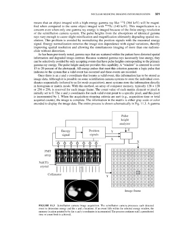
encoded to display the image data. The entire process is shown schematically in Fig. 11.3. A gamma
Pulse
height
analyzer
Energy Position Y Z
signal signals
X
X
PMT 0 0 0 0 0 0 0 0 0 0 0 0
0 0 0 0 0 0 0 0 0 0 0 0
array
0 0 0 0 0 0 0 0 0 0 0 0
0 0 0 0 0 0 0 0 0 0 0 0
NaI(Tl) 0 0 0 0 0 0 0 0 0 0 0 0
crystal 0 0 0 0 0 0 0 0 0 0 0 0
0 0 0 0 0 0 0 0 0 0 0 0
0 0 0 0 0 0 0 1 1 0 0 0 0
Collimator
Y 0 0 0 0 0 0 0 0 0 0 0 0
0 0 0 0 0 0 0 0 0 0 0 0
0 0 0 0 0 0 0 0 0 0 0 0
0 0 0 0 0 0 0 0 0 0 0 0
Image frame
FIGURE 11.3 Scintillation camera image acquisition. The scintillation camera processes each detected
event to determine energy and the x and y locations. If an event falls within the selected energy window, the
memory location pointed to by the x and y coordinates is incremented. The process continues until a preselected
time or count limit is achieved.