
Page 340 - Biomedical Engineering and Design Handbook Volume 2, Applications
P. 340
318 DIAGNOSTIC EQUIPMENT DESIGN
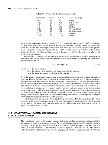
TABLE 11.1 Common Nuclear Medicine Radionuclides
Radionuclide Decay Half-life Gamma ray energy
99m Tc IT 6 h 140 keV
111 In EC 67 h 172, 247 keV
131 I β− 8 d 364 keV
123 I EC 13 h 159 keV
67 Ga EC 78 h 93, 185, 300 keV
201 Tl EC 73 h 60–80 keV (x-rays)
81m Kr IT 13 s 190 keV
133 Xe β− 5.3 d 80 keV
gamma rays makes detection more difficult. This is especially true for the 511-keV annihilation
photons associated with PET. As a result, the images produced in nuclear medicine studies are
much noisier and have worse spatial resolution than those generated from computed tomography
(CT) or magnetic resonance imaging (MRI). In order to appreciate these problems and how they
affect the design of nuclear medicine imaging devices, we will briefly review the physics of
gamma ray interactions.
The intensity of gamma rays traveling through material is gradually reduced by absorption or
scattering. This loss of gamma rays is referred to as attenuation and is described by the exponential
equation shown below:
I(x) = Io exp(−μx) (11.1)
where Io = the initial intensity
I(x) = the intensity after traveling a distance x through the material
μ= the linear attenuation coefficient of the material.
Over the range of gamma ray energies used in radionuclide imaging, the two primary interactions
that contribute to the attenuation coefficient are photoelectric absorption and Compton scattering.
Photoelectric absorption refers to the total absorption of the gamma ray by an inner-shell atomic
electron and is the primary interaction in high atomic number (Z) materials such as sodium iodide
(the detector material used in the scintillation camera) and lead. In low Z materials such as body tissues,
its contribution to attenuation is relatively small. Compton scattering occurs when the incoming
gamma ray interacts with a loosely bound outer shell electron. A portion of the gamma ray energy
is imparted to the electron and the remaining energy is left with the scattered photon. The amount of
energy lost in the event depends on the angle between the gamma ray and scattered photon. Compton
scattering is the dominant interaction in body tissues.
High attenuation is desirable in detecting and shielding materials. Ideally, materials used for these
purposes would absorb every gamma ray. In the body, attenuation is very undesirable since it reduces
the number of events that can be acquired and scattered radiation that reaches the detector causes a
significant loss of image contrast.
11.2 CONVENTIONAL GAMMA RAY IMAGING:
SCINTILLATION CAMERA
The scintillation camera is the primary imaging instrument used in conventional nuclear medicine
and is often referred to as a gamma camera. The scintillation camera is a position-sensitive gamma
ray imager. Although the entire field of view is available for detection, it processes one event at a
time. The spatial resolution is approximately 10 mm and it yields a count rate of 200 to 300 cpm/μCi
in the field of view. The field of view covers a large portion of the body and is typically 40 × 50 cm,