
Page 347 - Biomedical Engineering and Design Handbook Volume 2, Applications
P. 347
NUCLEAR MEDICINE IMAGING INSTRUMENTATION 325
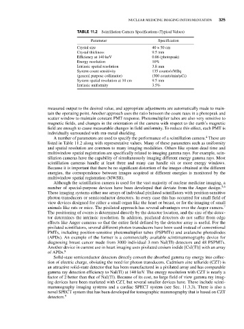
TABLE 11.2 Scintillation Camera Specifications (Typical Values)
Parameter Specification
Crystal size 40 × 50 cm
Crystal thickness 9.5 mm
Efficiency at 140 keV 0.86 (photopeak)
Energy resolution 10%
Intrinsic spatial resolution 3.8 mm
System count sensitivity 135 counts/s/MBq
(general purpose collimator) (300 counts/min/μCi)
System spatial resolution at 10 cm 9.5 mm
Intrinsic uniformity 3.5%
measured output to the desired value, and appropriate adjustments are automatically made to main-
tain the operating point. Another approach uses the ratio between the count rates in a photopeak and
scatter window to maintain constant PMT response. Photomultiplier tubes are also very sensitive to
magnetic fields, and changes in the orientation of the camera with respect to the earth’s magnetic
field are enough to cause measurable changes in field uniformity. To reduce this effect, each PMT is
individually surrounded with mu-metal shielding.
4
A number of parameters are used to specify the performance of a scintillation camera. These are
listed in Table 11.2 along with representative values. Many of these parameters such as uniformity
and spatial resolution are common to many imaging modalities. Others like system dead time and
multiwindow spatial registration are specifically related to imaging gamma rays. For example, scin-
tillation cameras have the capability of simultaneously imaging different energy gamma rays. Most
scintillation cameras handle at least three and many can handle six or more energy windows.
Because it is important that there be no significant distortion of the images obtained at the different
energies, the correspondence between images acquired at different energies is monitored by the
multiwindow spatial registration (MWSR).
Although the scintillation camera is used for the vast majority of nuclear medicine imaging, a
number of special-purpose devices have been developed that deviate from the Anger design. 5,6
These imaging systems either use arrays of individual pixilated scintillators with position-sensitive
photon transducers or semiconductor detectors. In every case this has occurred for small field of
view devices designed for either a small organ like the heart or breast, or for the imaging of small
animals like rats or mice. The pixilated approach has several advantages over the Anger camera. 7
The positioning of events is determined directly by the detector location, and the size of the detec-
tor determines the intrinsic resolution. In addition, pixilated detectors do not suffer from edge
effects like Anger cameras so that the entire field defined by the detector array is useful. For the
pixilated scintillators, several different photon transducers have been used instead of conventional
PMTs, including position-sensitive photomultiplier tubes (PSPMTs) and avalanche photodiodes
(APDs). An example of the former is a commercially available scintimammography device for
diagnosing breast cancer made from 3000 individual 3-mm NaI(Tl) detectors and 48 PSPMTs.
Another device in current use in heart imaging uses pixilated cesium iodide [CsI(Tl)] with an array
of APDs. 8
Solid-state semiconductor detectors directly convert the absorbed gamma ray energy into collec-
tion of electric charge, obviating the need for photon transducers. Cadmium zinc telluride (CZT) is
an attractive solid-state detector that has been manufactured in a pixilated array and has comparable
gamma ray detection efficiency to NaI(Tl) at 140 keV. The energy resolution with CZT is nearly a
factor of 2 better than that of NaI(Tl). Because of its cost, no large field of view gamma ray imag-
ing devices have been marketed with CZT, but several smaller devices have. These include scinti-
mammography imaging systems and a cardiac SPECT system (see Sec. 11.3.3). There is also a
novel SPECT system that has been developed for tomographic mammography that is based on CZT
detectors. 9