
Page 349 - Biomedical Engineering and Design Handbook Volume 2, Applications
P. 349
NUCLEAR MEDICINE IMAGING INSTRUMENTATION 327
a number of different approaches have been used to accomplish this, the most common method
moves the detectors radially in and out as a function of rotation angle. An example bone SPECT
study is shown in Fig. 11.7b.
The projection images acquired from a SPECT study do not reflect the line integrals of activity
within the patient. The primary reason for this is the attenuation of the internally emitted gamma
rays by body tissues. If projection images are reconstructed without correcting for tissue attenua-
tion, the resulting images will have artifacts. This is a significant problem with myocardial perfu-
sion since the attenuation artifacts can be mistaken for coronary artery disease. Accurate SPECT
attenuation correction requires an independent measurement of the tomographic attenuation coeffi-
cients for the volume being imaged. One solution for obtaining this information is to collect trans-
mission data through the patient from an external radioactive source using the gamma camera as a
11
crude CT scanner. Because of the multiple energy imaging capability of the scintillation camera, this
transmission information can be collected simultaneously as part of the SPECT acquisition so long as
the transmission source and the radiotracer have different gamma ray energies. Typically the radio-
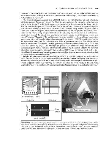
tracer is labeled with 99m Tc with a 140-keV gamma ray, while the transmission source is 153 Gd with
a 100-keV gamma ray (Fig. 11.8). Although the quality of the attenuation maps obtained for this
approach are poor, there is sufficient information to diminish the attenuation artifacts. An improved
attenuation correction can be obtained with a combined SPECT and CT device which will be dis-
cussed later. Attenuation compensation requires the use of an iterative reconstruction algorithm that
can incorporate the attenuation maps. 12
Despite the energy discrimination available on all SPECT systems, Compton scattered radiation
still accounts for about 30 to 40 percent of the acquired counts in SPECT imaging. Scattered radia-
tion not only decreases contrast, it also impacts other corrections. For example, when attenuation cor-
rection is applied without also correcting for scattered radiation, the count density in the heart walls
near the liver may be overenhanced. Scatter correction has been performed in several different ways, 13
Detector 1 Detector 1
Detector 2
100 keV
Collimated line source
End view Side view
Mask size (Y) 1 1 2 source Gd-153 Transmission source
Line
Mask width (X)
FIGURE 11.8 Transmission imaging with a radionuclide source. Attenuation correction is required for accurate SPECT.
In the thorax, this requires the acquisition of a transmission study. This shows one possible configuration where scanning
line sources of 153 Gd are translated across the field of view using the gamma camera as a crude CT scanner. This infor-
mation is used to correct myocardial perfusion studies for tissue attenuation. (Courtesy of GE Medical Systems.)