
Page 440 - Biomedical Engineering and Design Handbook Volume 2, Applications
P. 440
418 SURGERY
surgery) is removed. Then the femur is fixed to the base of the robot and a redundant position sensor
is attached to the bone to detect any slipping of the bone relative to the fixation device. Then a
3D digitizer is used to locate a number of points on the bone surface. These points are used to com-
pute the coordinate transformation between the robot and CT images used for planning and (thus) to
the patient’s bone. The surgeon then hand-guides the robot to an approximate initial position using
a force sensor mounted between the robot’s tool holder and the surgical cutter. The robot then cuts
the desired shape while monitoring cutting forces, bone motion, and other safety sensors. The
surgeon also monitors progress and can interrupt the robot at any time. If the procedure is paused for
any reason, there are a number of error recovery procedures available to permit the procedure to be
resumed or restarted at one of several defined checkpoints. Once the desired shape has been cut,
surgery proceeds manually in the normal manner. The procedural flow for robotic knee replacement
surgery is quite similar.
Robotically Assisted Percutaneous Therapy. One of the first uses of robots in surgery was posi-
tioning of needle guides in stereotactic neurosurgery. 24,108,109 This is a natural application, since the
skull provides a rigid frame of reference. However, the potential application of localized therapy is
much broader. Percutaneous therapy fits naturally within the broader paradigm of surgical CAD/CAM
systems. The basic process involves planning a patient-specific therapy pattern, delivering the ther-
apy through a series of percutaneous access steps, assessing what was done, and using this feedback
to control therapy at several time scales. The ultimate goal of current research is to develop systems
that execute this process with robotic assistance under a variety of widely available and deployable
image modalities, including ultrasound, x-ray fluoroscopy, and conventional MRI and CT scanners.
Current work at Johns Hopkins University is typical of this activity. Our approach has emphasized
the use of “remote center-of-motion” (RCM) manipulators to position needle guides under real-time
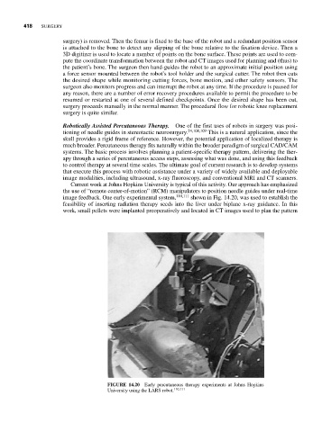
image feedback. One early experimental system, 110,111 shown in Fig. 14.20, was used to establish the
feasibility of inserting radiation therapy seeds into the liver under biplane x-ray guidance. In this
work, small pellets were implanted preoperatively and located in CT images used to plan the pattern
FIGURE 14.20 Early percutaneous therapy experiments at Johns Hopkins
University using the LARS robot. 110,111