
Page 441 - Biomedical Engineering and Design Handbook Volume 2, Applications
P. 441
COMPUTER-INTEGRATED SURGERY AND MEDICAL ROBOTICS 419
of therapy seeds. The fiducial pellets were relocated in the biplane x-rays and used to register the pre-
operative plan to a modified LARS robot 112,113 used to implant the treatment seeds. Although this
experiment and related work directed at placing needles into the kidney 114,115 established the basic
feasibility of our approach, we concluded that significant improvements in the robot would be needed.
Subsequent work has focused on development of a modular family of very compact component
subsystems and end effectors that could be configured for use in a variety of imaging and surgical
environments. Figure 14.9 shows a novel RCM linkage with a radiolucent needle driver (“PAKY”)
developed by Stoianovici et al. that forms a key component in this next generation system. Figure 14.11
shows the RCM device with a novel end-effector developed by Susil and Masamune that permits the
computer to determine the needle pose to be computed with respect to a CT or MRI scanner using a
single image slice. 42,44,45 This arrangement can have significant advantages in reducing setup costs and
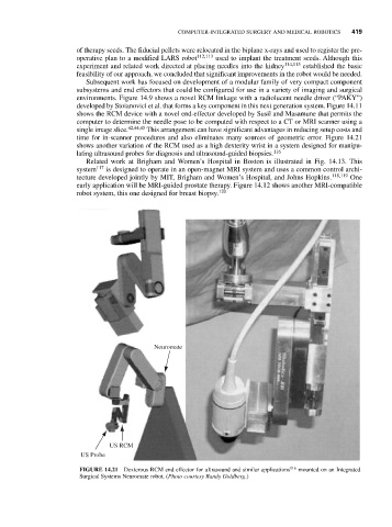
time for in-scanner procedures and also eliminates many sources of geometric error. Figure 14.21
shows another variation of the RCM used as a high dexterity wrist in a system designed for manipu-
lating ultrasound probes for diagnosis and ultrasound-guided biopsies. 116
Related work at Brigham and Women’s Hospital in Boston is illustrated in Fig. 14.13. This
system 117 is designed to operate in an open-magnet MRI system and uses a common control archi-
tecture developed jointly by MIT, Brigham and Women’s Hospital, and Johns Hopkins. 118,119 One
early application will be MRI-guided prostate therapy. Figure 14.12 shows another MRI-compatible
robot system, this one designed for breast biopsy. 120
Neuromate
US RCM
US Probe
FIGURE 14.21 Dexterous RCM end effector for ultrasound and similar applications 116 mounted on an Integrated
Surgical Systems Neuromate robot. (Photo courtesy Randy Goldberg.)