
Page 446 - Biomedical Engineering and Design Handbook Volume 2, Applications
P. 446
424 SURGERY
14.6.2 Image Processing, Visualization, and Modeling
Medical image processing and visualization have greatly benefited from the increase in speed and
performance of standard computing equipment. Specialized automatic and semiautomatic segmen-
tation techniques for an ever-increasing number of image modalities and anatomical structures have
been developed over the past 5 years. 171 The most commonly studied anatomical structures include
the brain and its vasculature, the heart, the liver, the colon, and bones. Three-dimensional visualiza-
tion is gaining acceptance in the clinical environment, especially for the visualization of complex
anatomical structures and pathologies. A current trend is patient-specific modeling for preoperative
planning and interventional procedures.
14.6.3 Preoperative Analysis, Planning, and Registration
Preoperative analysis and planning has remained very much procedure and equipment-specific. It is
usually incorporated within the systems (Fig. 14.26c). Work on registration has emphasized multimodal
and nonrigid registration, incorporating real-time laparoscopic video and ultrasound images. 171
A B
C
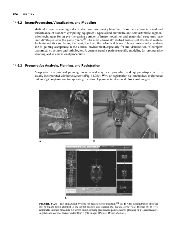
FIGURE 14.26 The SpineAssist System for pedicle screw insertion. 172 (a) In vitro demonstration showing
the miniature robot clamped to the spinal process and guiding the pedicle screw hole drilling; (b) in vivo
minimally invasive procedure; (c) screen dump showing preoperative pedicle screws planning on CT axial (center),
sagittal, and coronal (center and bottom right) images (Photos: Moshe Shoham).