
Page 249 - Biomimetics : Biologically Inspired Technologies
P. 249
Bar-Cohen : Biomimetics: Biologically Inspired Technologies DK3163_c008 Final Proof page 235 21.9.2005 3:08am
Molecular Design of Biological and Nano-Materials 235
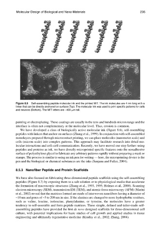
Figure 8.6 Self-assembling peptide molecular ink and the printed MIT. The ink molecules are 4 nm long with a
linker that can be directly anchored on surface (Top). The molecular ink was used to print specific patterns for cells
and neurons (Bottom). The MIT letters are ~400 mm tall.
painting or electroplating. These coatings are usually in the tens and hundreds micron range and the
interface is often not complementary at the molecular level. Thus, erosion is common.
We have developed a class of biologically active molecular ink (Figure 8.6), self-assembling
peptides with linkers that anchor on surfaces (Zhang et al., 1999). In conjunction with self-assembled
monolayers prepared through microcontact printing, we can place molecules (nanometer scale) and
cells (micron scale) into complex patterns. This approach may facilitate research into detail mo-
lecular interactions and cell–cell communication. Recently, we have moved one step further: using
peptides and proteins as ink, we have directly microprinted specific features onto the nonadhesive
surface of polyethylene glycol to fabricate any arbitrary patterns rapidly without preparing a mask or
stamps. The process is similar to using an ink pen for writing — here, the microprinting device is the
pen and the biological or chemical substances are the inks (Sanjana and Fuller, 2004).
8.5.3 Nanofiber Peptide and Protein Scaffolds
We have also focused on fabricating three-dimensional peptide scaffolds using the self-assembling
peptides (Figure 8.7) by exposing them to a salt solution or to physiological media that accelerate
the formation of macroscopic structures (Zhang et al., 1993, 1995; Holmes et al., 2000). Scanning
electron microscopy (SEM), transmission EM (TEM), and atomic force microscopy (AFM) (Marini
et al., 2002) reveal that the matrices formed are made of interwoven nanofibers having a diameter of
~10 nm and pores of ~5 to 200 nm in size. If the alanines are changed to more hydrophobic residues,
such as valine, leucine, isoleucine, phenylalanine, or tyrosine, the molecules have a greater
tendency to self-assemble and form peptide matrices. These simple, defined and tailor-made self-
assembling peptides have provided the first de novo designed scaffolds for three-dimensional cell
culture, with potential implications for basic studies of cell growth and applied studies in tissue
engineering and ultimately regenerative medicine (Kisiday et al., 2002; Zhang, 2004).