
Page 251 - Biomimetics : Biologically Inspired Technologies
P. 251
Bar-Cohen : Biomimetics: Biologically Inspired Technologies DK3163_c008 Final Proof page 237 21.9.2005 3:08am
Molecular Design of Biological and Nano-Materials 237
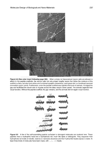
Figure 8.8 (See color insert following page 302) When primary rat hippocampal neuron cells are allowed to
attach to the peptide scaffolds, the neuron cells not only project lengthy axons that follow the contours of the
scaffold surface, but also form active and functional synaptic connections, each green dot is a functional neuronal
connection (upper panel). Furthermore, when the peptide scaffold was injected into brain of animals, it bridged the
gap and facilitated the neural cells to migrate across the deep canyon (lower panel). The animals regained their
visual function. Without the peptide scaffold, the gap remains, and the animals did not regain visual function.
Figure 8.9 A few of the self-assembling peptide surfactant or detergent molecules are modeled here. These
peptides have a hydrophilic head and a hydrophobic tail, much like lipids or detergents. They sequester their
hydrophobic tail inside of micelle, vesicles or nanotube structures and their hydrophilic heads expose to water. At
least three kinds of molecules have been made, with , þ, /þ heads.