
Page 61 - Biomimetics : Biologically Inspired Technologies
P. 61
Bar-Cohen : Biomimetics: Biologically Inspired Technologies DK3163_c002 Final Proof page 47 21.9.2005 11:39am
Biomimetics of Muscle Design 47
with the cell membrane via specialized structural proteins like desmin (Figure 2.1). Based on the
evidence from animal experiments (Huijing, 1999), it is now thought that the force of individual
sarcomeres finds its way to the outside via both serial and parallel pathways.
2.4 MUSCLE DESIGN
Within the animal kingdom, the variety in muscle designs is stunning. There are bulky muscles (m.
gluteus maximus), long slender muscles (sartorius), muscles with short fibers attached to long
tendons (m. gastrocnemius), pennate muscles, etc. Muscle design is highly variable within an
animal and also between species. It appears as if there is a specialized muscle design for each
possible function (Otten, 1988). It is beyond the scope of this chapter to review all possible designs
and functions, and therefore a few basic design principles of muscle will be discussed. Muscles are
built from sarcomeres and as a consequence it has two basic design options to tune into functional
demands. It can modify either the design or the arrangement of the sarcomeres. Both options appear
to have been explored by Nature.
2.4.1 Not all Sarcomeres Are Alike
Invertebrates appear to have explored the possibilities of sarcomere design to its full potential.
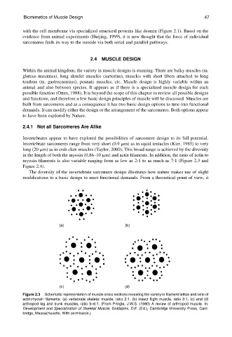
Invertebrate sarcomeres range from very short (0.9 mm) as in squid tentacles (Kier, 1985) to very
long (20 mm) as in crab claw muscles (Taylor, 2000). This broad range is achieved by the diversity
in the length of both the myosin (0.86–10 mm) and actin filaments. In addition, the ratio of actin to
myosin filaments is also variable ranging from as low as 2:1 to as much as 7:1 (Figure 2.3 and
Figure 2.4).
The diversity of the invertebrate sarcomere design illustrates how nature makes use of slight
modifications to a basic design to meet functional demands. From a theoretical point of view, it
Figure 2.3 Schematic representation of muscle cross sections revealing the variety in filament lattice and ratio of
actin:myosin filaments: (a) vertebrate skeletal muscle, ratio 2:1, (b) insect flight muscle, ratio 3:1, (c) and (d)
arthropod leg and trunk muscles, ratio 5–6:1. (From Pringle, J.W.S. (1980) A review of arthropod muscle. In:
Development and Specialization of Skeletal Muscle, Goldspink, D.F. (Ed.), Cambridge University Press, Cam-
bridge, Massachusetts. With permission.)