
Page 363 - Carrahers_Polymer_Chemistry,_Eighth_Edition
P. 363
326 Carraher’s Polymer Chemistry
10.2.1 PRIMARY STRUCTURE
The term primary structure is used to describe the sequence of amino acid units (confi guration) in
a polypeptide chain. Thus, Equation 10.5 describes a primary structure.
10.2.2 SECONDARY STRUCTURE
The term secondary structure is used to describe the molecular shape or conformation of a mole-
cule. The most important factor in determining the secondary structure of materials is its precise
structure. For proteins, it is the amino acid sequence. Hydrogen bonding is also an important factor
in determining the secondary structures of natural materials and those synthetic materials that can
hydrogen bond. In fact, for proteins, secondary structures are generally those that allow a maximum
amount of hydrogen bonding. This hydrogen bonding also acts to stabilize the secondary structure
while cross-linking acts to lock-in a structure.
In nature, the two most common secondary structures are helices and sheets. In nature, extended
helical conformations appear to be utilized in two major ways: to provide linear systems for the
storage, duplication, and transmission of information (DNA, RNA), and to provide inelastic fi bers
for the generation and transmission of forces (F-actin, myosin, and collagen). Examples of the var-
ious helical forms found in nature are single helix (messenger and ribosomal DNA), double helix
(DNA), triple helix (collagen fibrils), and complex multiple helices (myosin). Generally, these single
and double helices are readily soluble in dilute aqueous solution. Often solubility is only achieved
after the inter- and intrahydrogen bonding is broken.
There are a variety of examples in which linear or helical polypeptide chains are arranged in par-
allel rows. The two major forms that exist for proteins are illustrated in Figures 10.2 through 10.5. The
chains can have the N→C directions running parallel making a parallel beta sheet (Figure 10.4), or
they can have the N→C directions running antiparallel giving antiparallel beta sheets (Figure 10.5).
The structure of proteins generally fall into three groupings—fibers, membrane, and globular.
The structural proteins such as the keratines, collagen, and the elastin are largely fibrous. A reoc-
curring theme with respect to conformation is that the preferential secondary structures of fi brous
synthetic and natural polymers approximates that of a pleated sheet (or skirt) or helix. The pleated
sheet structures in proteins are referred to as beta arrangements (Figure 10.5). In general, proteins
with bulky groups take on a helical secondary structure while those with less bulky groups exist as
beta sheets.
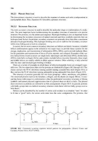
Helices can be described by the number of amino acid residues in a complete “turn.” In order
to fit into a “good” helix, the amino acids must have the same configuration. For proteins, that
Alpha-helix
2 7 Ribbon
H H
O O O
N N R
R N N N
O O O
H H H
3 10 Helix
Pi Helix
FIGURE 10.2 Commonly occurring repetitive helical patterns for polypeptides. (Source: Coates and
Carraher, Polymer News, 9(3):77 (1983)).
9/14/2010 3:41:09 PM
K10478.indb 326 9/14/2010 3:41:09 PM
K10478.indb 326