
Page 581 - Carrahers_Polymer_Chemistry,_Eighth_Edition
P. 581
544 Carraher’s Polymer Chemistry
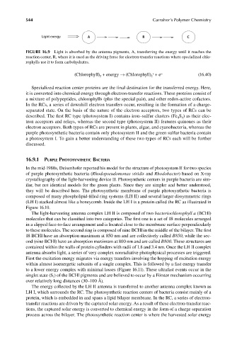
Light energy A R C
FIGURE 16.9 Light is absorbed by the antenna pigments, A, transferring the energy until it reaches the
reaction center, R, where it is used as the driving force for electron-transfer reactions where specialized chlo-
rophylls use it to form carbohydrates.
+
(Chlorophyll) + energy → (Chlorophyll) + e − (16.40)
2
2
Specialized reaction center proteins are the final destination for the transferred energy. Here,
it is converted into chemical energy through electron-transfer reactions. These proteins consist of
a mixture of polypeptides, chlorophylls (plus the special pair), and other redox-active cofactors.
In the RCs, a series of downhill electron transfers occur, resulting in the formation of a charge-
separated state. On the basis of the nature of the electron acceptors, two types of RCs can be
described. The first RC type (photosystem I) contains iron–sulfur clusters (Fe S ) as their elec-
4 4
tron acceptors and relays, whereas the second type (photosystem II) features quinones as their
electron acceptors. Both types of RCs are present in plants, algae, and cyanobacteria, whereas the
purple photosynthetic bacteria contain only photosystem II and the green sulfur bacteria contain
a photosystem I. To gain a better understanding of these two types of RCs each will be further
discussed.
16.9.1 PURPLE PHOTOSYNTHETIC BACTERIA
In the mid-1980s, Deisenhofer reported his model for the structure of photosystem II for two species
of purple photosynthetic bacteria (Rhodopseudomonas viridis and Rhodobacter) based on X-ray
crystallography of the light-harvesting device II. Photosynthetic centers in purple bacteria are sim-
ilar, but not identical models for the green plants. Since they are simpler and better understood,
they will be described here. The photosynthetic membrane of purple photosynthetic bacteria is
composed of many phospholipid-filled ring systems (LH II) and several larger dissymmetric rings
(LH I) stacked almost like a honeycomb. Inside the LH I is a protein called the RC as illustrated in
Figure 16.10.
The light-harvesting antenna complex LH II is composed of two bacteriochlorophyll a (BCHl)
molecules that can be classified into two categories. The first one is a set of 18 molecules arranged
in a slipped face-to-face arrangement and is located close to the membrane surface perpendicularly
to these molecules. The second ring is composed of nine BCHl in the middle of the bilayer. The fi rst
18 BCHl have an absorption maximum at 850 nm and are collectively called B850, while the sec-
ond (nine BCHl) have an absorption maximum at 800 nm and are called B800. These structures are
contained within the walls of protein cylinders with radii of 1.8 and 3.4 nm. Once the LH II complex
antenna absorbs light, a series of very complex nonradiative photophysical processes are triggered.
First the excitation energy migrates via energy transfers involving the hopping of excitation energy
within almost isoenergetic subunits of a single complex. This is followed by a fast energy transfer
to a lower energy complex with minimal losses (Figure 16.11). These ultrafast events occur in the
singlet state (S ) of the BCHl pigments and are believed to occur by a Förster mechanism occurring
1
over relatively long distances (30–100 Å).
The energy collected by the LH II antenna is transferred to another antenna complex known as
LH I, which surrounds the RC. The photosynthetic reaction centers of bacteria consist mainly of a
protein, which is embedded in and spans a lipid bilayer membrane. In the RC, a series of electron-
transfer reactions are driven by the captured solar energy. As a result of these electron-transfer reac-
tions, the captured solar energy is converted to chemical energy in the form of a charge separation
process across the bilayer. The photosynthetic reaction center is where the harvested solar energy
9/14/2010 3:43:15 PM
K10478.indb 544 9/14/2010 3:43:15 PM
K10478.indb 544