
Page 231 - Color Atlas of Biochemistry
P. 231
222 Organelles
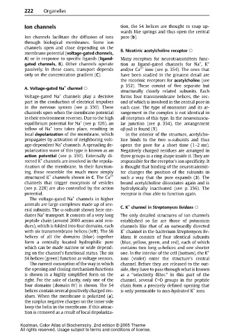
Ion channels tion, the S4 helices are thought to snap up-
wardslike springsand thus open the central
Ion channels facilitate the diffusion of ions pore (b).
through biological membranes. Some ion
channels open and close depending on the
membrane potential (voltage-gated channels, B. Nicotinic acetylcholine receptor
A) or in response to specific ligands (ligand- Many receptors for neurotransmitters func-
+
gated channels, B). Other channels operate tion as ligand-gated channels for Na ,K +
passively. In these cases, transport depends and/or Ca 2+ ions (see p. 354). The ones that
only on the concentration gradient (C). have been studied in the greatest detail are
the nicotinic receptors for acetylcholine (see
p. 352). These consist of five separate but
+
A. Voltage-gated Na channel
structurally closely related subunits. Each
+
Voltage-gated Na channels play a decisive forms four transmembrane helices, the sec-
part in the conduction of electrical impulses ond of which is involved in the central pore in
in the nervous system (see p. 350). These each case. The type of monomer and its ar-
channels open when the membrane potential rangement in the complex is not identical in
in their environment reverses. Due to the high all receptors of this type. In the neuromuscu-
+
equilibrium potential for Na (see p. 126), an lar junction (see p. 334), the arrangement
+
inflow of Na ions takes place, resulting in αβγαδ is found (1).
local depolarization of the membrane, which In the interior of the structure, acetylcho-
propagates by activation of neighboring volt- line binds to the two α-subunits and thus
+
age-dependent Na channels. A spreading de- opens the pore for a short time (1–2 ms).
polarization wave of this type is known as an Negatively charged residues are arranged in
action potential (see p. 350). Externally di- threegroups in a ring shapeinsideit. They are
+
rected K channels are involved in the repola- responsible for the receptor’s ion specificity. It
rization of the membrane. In their function- is thought that binding of the neurotransmit-
ing, these resemble the much more simply ter changes the position of the subunits in
+
structured K channels shown in C.The Ca 2+ such a way that the pore expands (3). The
channels that trigger exocytosis of vesicles bound acetylcholine dissociates again and is
(see p. 228) are also controlled by the action hydrolytically inactivated (see p. 356). The
potential. receptor is thus able to function again.
+
The voltage-gated Na channels in higher
animals are large complexes made up of sev- +
eral subunits. The α-subunit shown here me- C. K channel in Streptomyces lividans
+
diates Na transport. It consists of a very long The only detailed structures of ion channels
peptide chain (around 2000 amino acid resi- established so far are those of potassium
dues), which is folded into four domains, each channels like that of an outwardly directed
+
with six transmembrane helices (left). The S6 K channel in the bacterium Streptomyces liv-
helices of all the domains (blue) together idans. It consists of four identical subunits
form a centrally located hydrophilic pore (blue, yellow, green, and red), each of which
which can be made narrow or wide depend- contains two long α-helices and one shorter
ing on the channel’s functional status. The six one. In the interior of the cell (bottom), the K +
S4 helices (green) function as voltage sensors. ions (violet) enter the structure’s central
The current conception of the way in which channel. Before they are released to the out-
the opening and closing mechanism functions side, they have to pass through what is known
is shown in a highly simplified form on the as a “selectivity filter.” In this part of the
right. For the sake of clarity, only one of the channel, several C=O groups in the peptide
four domains (domain IV) is shown. The S4 chain form a precisely defined opening that
+
helices contain several positively charged res- is only permeable to non-hydrated K ions.
idues. When the membrane is polarized (a),
the surplus negative charges on the inner side
keep the helix in the membrane. If this attrac-
tion is removed as a result of local depolariza-
Koolman, Color Atlas of Biochemistry, 2nd edition © 2005 Thieme
All rights reserved. Usage subject to terms and conditions of license.