
Page 245 - Computational Modeling in Biomedical Engineering and Medical Physics
P. 245
234 Computational Modeling in Biomedical Engineering and Medical Physics
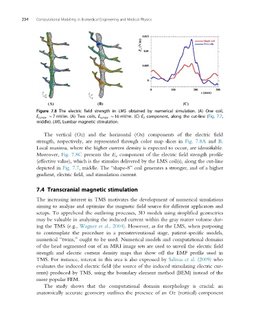
Figure 7.8 The electric field strength in LMS obtained by numerical simulation. (A) One coil,
E z,max B7 mV/m. (A) Two coils, E x,max B16 mV/m. (C) E z component, along the cut-line (Fig. 7.7,
middle). LMS, Lumbar magnetic stimulation.
The vertical (Oz) and the horizontal (Ox) components of the electric field
strength, respectively, are represented through color map slices in Fig. 7.8A and B.
Local maxima, where the higher current density is expected to occur, are identifiable.
Moreover, Fig. 7.8C presents the E z component of the electric field strength profile
(effective value), which is the stimulus delivered by the LMS coil(s), along the cut-line
depicted in Fig. 7.7, middle. The “shape-8” coil generates a stronger, and of a higher
gradient, electric field, and stimulation current.
7.4 Transcranial magnetic stimulation
The increasing interest in TMS motivates the development of numerical simulations
aiming to analyze and optimize the magnetic field source for different applicators and
setups. To apprehend the outlining processes, 3D models using simplified geometries
may be valuable in analyzing the induced current within the gray matter volume dur-
ing the TMS (e.g., Wagner et al., 2004). However, as for the LMS, when purposing
to contemplate the procedure in a preinterventional stage, patient-specific models,
numerical “twins,” ought to be used. Numerical models and computational domains
of the head segmented out of an MRI image sets are used to unveil the electric field
strength and electric current density maps that show off the EMF profile used in
TMS. For instance, interest in this area is also expressed by Salinas et al. (2009) who
evaluates the induced electric field (the source of the induced stimulating electric cur-
rents) produced by TMS, using the boundary element method (BEM) instead of the
more popular FEM.
The study shows that the computational domain morphology is crucial: an
anatomically accurate geometry outlines the presence of an Oz (vertical) component