
Page 247 - Computational Modeling in Biomedical Engineering and Medical Physics
P. 247
236 Computational Modeling in Biomedical Engineering and Medical Physics
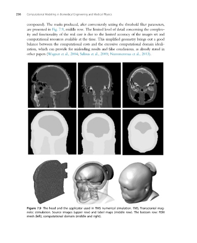
compound). The masks produced, after conveniently setting the threshold filter parameters,
are presented in Fig. 7.9, middle row. The limited level of detail concerning the complex-
ity and functionality of the real case is due to the limited accuracy of the images set and
computational resources available at the time. This simplified geometry brings out a good
balance between the computational costs and the excessive computational domain ideali-
zation, which can provide for misleading results and false conclusions, as already stated in
other papers (Wagner et al., 2004; Salinas et al., 2009; Nummenmaa et al., 2013).
Figure 7.9 The head and the applicator used in TMS numerical simulation. TMS, Transcranial mag-
netic stimulation. Source images (upper row) and label maps (middle row). The bottom row: FEM
mesh (left), computational domain (middle and right).