
Page 251 - Computational Modeling in Biomedical Engineering and Medical Physics
P. 251
240 Computational Modeling in Biomedical Engineering and Medical Physics
this possible side effect of the MFT when applied, for example, for postsurgical healing of a
fractured femur shaft. This kind of fracture is a severe handicap, the associated recovery lasts
and it is accompanied by immobilization and discomfort. The specific treatment is, in general,
surgical and purposed to stabilize the fractured bone with metallic structures. Plates and screws
outside the bone or an external fixator are the common solutions (Femur, 2020; Uptodate,
2020) but, in severe cases, a rod is introduced in the center of the medullar canal and fastened
with screws (Su et al., 2015). Intensive numerical modeling is devoted, in the presurgical
phase, to predict the structural stability and dynamic response of such consolidated structures
under various solicitations, mainly mechanical (e.g., Coquim et al., 2018).
The unavoidable immobilization rapidly leads to the depreciation of the muscle,
the reduction of its volume and its physical fitness, and even musculoskeletal and neu-
ronal disorder can occur. Physiotherapy may help speeding up restoring mobility, and
one of the most popular and successful is MFT—used also in orthopedics and rheuma-
tology or for the treatment of internal diseases. MFT has been proven to help relieve
pain and accelerate the healing time. It has an important influence on trophic stimula-
tion of collagen and bones by producing microcurrents that speed up osteogenesis
(èada-Tondrya, 2019; Baerov et al., 2020; Efisioterapia.Net, 2020).
We touch also the possible amplification of the side effects of the MFT due to the
electromagnetic heating of metallic implants that are the siege of induced currents.
Numerical simulations are conducted using computational models that represent the
anatomy of the upper leg and an orthopedic implanted device that is exposed to a har-
monic magnetic field—first a sketchy 3D CAD construct and then a medical images-
based reconstructed domain.
Modeling the magnetic field therapy
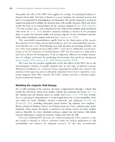
For a CAD rendering of the anatomy, the tissue is approximated through a cylinder that
models the soft tissues, which hosts another cylinder that represents the femur, Fig. 7.12,
left. Stainless steel and titanium plates are usually used (Sahoo et al., 1994) but, recently,
there is a growing in using platinum in medical applications due to biocompatibility, inert-
ness within the body, durability, electrical conductivity, and radiopacity (Cowley and
Woodward, 2011), including orthopedics (spinal fixation; hip implants, knee implants—
Biomet, Johnson & Johnson, Stryker, and Zimmer producers). Here, a platinum plate (paral-
lelepiped), which mimics the fixator, is attached to the femur—screws and other ancillary
parts are discarded. An outer cylindrical enclosure, capped (top and bottom) with infinite
elements subdomains, contains the anatomic volumes and closes the MF.
Once the outlining MFT processes are evidenced using this CAD construct, a real-
istic model is essential to achieve consistent results for medical applications to add
predictability and enhance the diagnosis accuracy. To this aim, reconstruction