
Page 248 - Computational Modeling in Biomedical Engineering and Medical Physics
P. 248
Magnetic stimulation and therapy 237
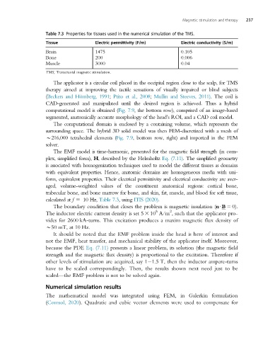
Table 7.3 Properties for tissues used in the numerical simulation of the TMS.
Tissue Electric permittivity (F/m) Electric conductivity (S/m)
Brain 1475 0.105
Bone 200 0.006
Muscle 3000 0.04
TMS, Transcranial magnetic stimulation.
The applicator is a circular coil placed in the occipital region close to the scalp, for TMS
therapy aimed at improving the tactile sensations of visually impaired or blind subjects
(Beckers and Hömberg, 1991; Ptito et al., 2008; Mullin and Steeves, 2011). The coil is
CAD-generated and manipulated until the desired region is achieved. Thus a hybrid
computational model is obtained (Fig. 7.9, the bottom row), comprised of an image-based
segmented, anatomically accurate morphology of the head’s ROI, and a CAD coil model.
The computational domain is enclosed by a containing volume, which represents the
surrounding space. The hybrid 3D solid model was then FEM-discretized with a mesh of
B216,000 tetrahedral elements (Fig. 7.9, bottom row, right) and imported in the FEM
solver.
The EMF model is time-harmonic, presented for the magnetic field strength (in com-
plex, simplified form), H, described by the Helmholtz Eq. (7.11). The simplified geometry
is associated with homogenization techniques used to model the different tissues as domains
with equivalent properties. Hence, anatomic domains are homogeneous media with uni-
form, equivalent properties. Their electrical permittivity and electrical conductivity are aver-
aged, volume-weighted values of the constituent anatomical regions: cortical bone,
trabecular bone, and bone marrow for bone, and skin, fat, muscle, and blood for soft tissue,
calculated at f 5 10 Hz, Table 7.3,using ITIS (2020).
The boundary condition that closes the problem is magnetic insulation (nUB 5 0).
2
5
The inductor electric current density is set 5 3 10 A/m , such that the applicator pro-
vides for 2600 kA-turns. This excitation produces a maxim magnetic flux density of
B50 mT, at 10 Hz.
It should be noted that the EMF problem inside the head is here of interest and
not the EMF, heat transfer, and mechanical stability of the applicator itself. Moreover,
because the PDE Eq. (7.11) presents a linear problem, its solution (the magnetic field
strength and the magnetic flux density) is proportional to the excitation. Therefore if
other levels of stimulation are acquired, say 1 1.5 T, then the inductor ampere-turns
have to be scaled correspondingly. Then, the results shown next need just to be
scaled—the EMF problem is not to be solved again.
Numerical simulation results
The mathematical model was integrated using FEM, in Galerkin formulation
(Comsol, 2020). Quadratic and cubic vector elements were used to compensate for