
Page 253 - Computational Modeling in Biomedical Engineering and Medical Physics
P. 253
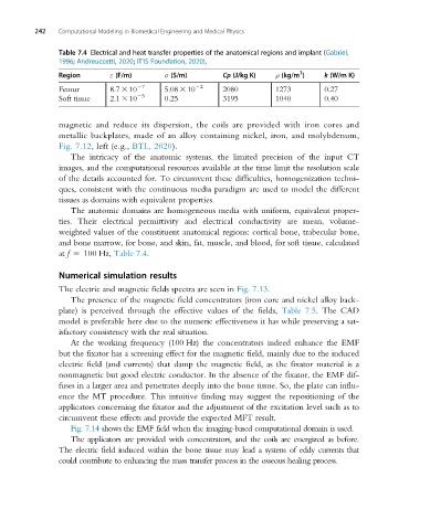
242   Computational Modeling in Biomedical Engineering and Medical Physics


                Table 7.4 Electrical and heat transfer properties of the anatomical regions and implant (Gabriel,
                1996; Andreuccetti, 2020; IT'IS Foundation, 2020).
                                                                            3
                Region       ε (F/m)      σ (S/m)        Cp (J/kg K)  ρ (kg/m )   k (W/m K)
                Femur        8.7 3 10 27  5.08 3 10 22   2080         1273        0.27
                Soft tissue  2.1 3 10 25  0.25           3195         1040        0.40


                magnetic and reduce its dispersion, the coils are provided with iron cores and
                metallic backplates, made of an alloy containing nickel, iron, and molybdenum,
                Fig. 7.12, left (e.g., BTL, 2020).
                   The intricacy of the anatomic systems, the limited precision of the input CT
                images, and the computational resources available at the time limit the resolution scale
                of the details accounted for. To circumvent these difficulties, homogenization techni-
                ques, consistent with the continuous media paradigm are used to model the different
                tissues as domains with equivalent properties.
                   The anatomic domains are homogeneous media with uniform, equivalent proper-
                ties. Their electrical permittivity and electrical conductivity are mean, volume-
                weighted values of the constituent anatomical regions: cortical bone, trabecular bone,
                and bone marrow, for bone, and skin, fat, muscle, and blood, for soft tissue, calculated
                at f 5 100 Hz, Table 7.4.


                Numerical simulation results
                The electric and magnetic fields spectra are seen in Fig. 7.13.
                   The presence of the magnetic field concentrators (iron core and nickel alloy back-
                plate) is perceived through the effective values of the fields, Table 7.5. The CAD
                model is preferable here due to the numeric effectiveness it has while preserving a sat-
                isfactory consistency with the real situation.
                   At the working frequency (100 Hz) the concentrators indeed enhance the EMF
                but the fixator has a screening effect for the magnetic field, mainly due to the induced
                electric field (and currents) that damp the magnetic field, as the fixator material is a
                nonmagnetic but good electric conductor. In the absence of the fixator, the EMF dif-
                fuses in a larger area and penetrates deeply into the bone tissue. So, the plate can influ-
                ence the MT procedure. This intuitive finding may suggest the repositioning of the
                applicators concerning the fixator and the adjustment of the excitation level such as to
                circumvent these effects and provide the expected MFT result.
                   Fig. 7.14 shows the EMF field when the imaging-based computational domain is used.
                   Theapplicators areprovidedwithconcentrators, and the coils are energized as before.
                The electric field induced within the bone tissue may lead a system of eddy currents that
                could contribute to enhancing the mass transfer process in the osseous healing process.