
Page 274 - Computational Modeling in Biomedical Engineering and Medical Physics
P. 274
Hyperthermia and ablation 263
A key observation is that image-based reconstruction techniques based on the
patient’s relevant medical scans (DICOM sets), for example (Morega et al., 2010), for
the computational domains, are required to implement the GHT as a convenient,
cost-efficient, noninvasive tool that may contribute to better understand the RFA pro-
cedure, and to its optimization.
Some thermographic considerations
Pennes model belongs to the “two-temperatures” thermal model, with the observa-
tion that one of the temperatures (blood) is a reference and not the result of a
convective-diffusion process. The benefits and limitations of this bioheat transfer
model were discussed in Chapter 1: Physical, Mathematical, and Numerical Modeling.
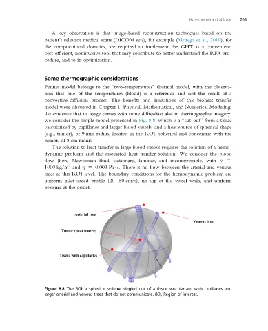
To evidence that its usage comes with some difficulties also in thermographic imagery,
we consider the simple model presented in Fig. 8.8, which is a “cut-out” from a tissue
vascularized by capillaries and larger blood vessels, and a heat source of spherical shape
(e.g., tumor), of 9 mm radius, located in the ROI, spherical and concentric with the
tumor, of 8 cm radius.
The solution to heat transfer in large blood vessels requires the solution of a hemo-
dynamic problem and the associated heat transfer solution. We consider the blood
flow (here Newtonian fluid) stationary, laminar, and incompressible, with ρ 5
3
1000 kg/m and η 5 0.003 Pa s. There is no flow between the arterial and venous
trees at this ROI level. The boundary conditions for the hemodynamic problem are
uniform inlet speed profile (20 50 cm/s), no-slip at the vessel walls, and uniform
pressure at the outlet.
Arterial tree
Venous tree
Tumor (heat source)
Tissue with capillaries
Figure 8.8 The ROI: a spherical volume singled out of a tissue vascularized with capillaries and
larger arterial and venous trees that do not communicate. ROI, Region of interest.