
Page 282 - Computational Modeling in Biomedical Engineering and Medical Physics
P. 282
Hyperthermia and ablation 271
FEM modeling media (Comsol, 2010 2019) may provide the solution of coupled
problems of different physical content with controllable numerical accuracy. The elec-
tromagnetic time constants are much smaller than the thermal and flow ones; thus,
Q emf , which is a result of the radiation problem, represents a stationary (r.m.s.) heat
source. The heat transfer and the hemodynamic flow in large arteries are one-way
coupled (Morega et al., 2014).
Thermal analysis in mild hyperthermia of soft tissue
An example of efficiently heating the target volume of tissue for moderate hyperther-
mia presents the case of a two pins applicator inserted in soft tissue, with the physical
properties of the liver. The physical and mathematical models were presented above,
and a comparison between the two cooling mechanisms through blood flow (by the
capillary network versus large vessels) is performed. Each antenna follows the model
shown in Fig. 8.11 (left), and the physical properties of the model with liver desig-
nated as target tissue are given in Table 8.5.
Homogeneous tissue model, with the vascularization provided by a network of capil-
lary blood vessels, is first analyzed, based on the Eqs. (8.10) and (8.11a). The optimal dis-
tance of 12 mm between the two pins was determined after a parametric study, following
an efficiency criterium: maximum volume of tissue heated as uniform as possible within
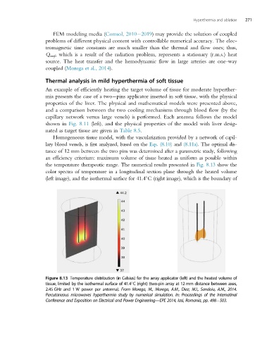
the temperature therapeutic range. The numerical results presented in Fig. 8.13 show the
color spectra of temperature in a longitudinal section plane through the heated volume
(left image), and the isothermal surface for 41.4 C (right image), which is the boundary of
Figure 8.13 Temperature distribution (in Celsius) for the array applicator (left) and the heated volume of
tissue, limited by the isothermal surface of 41.4 C (right) (two-pin array at 12 mm distance between axes,
2.45 GHz and 1 W power per antenna). From Morega, M., Morega, A.M., Diaz, M.I., Sandoiu, A.M., 2014.
Percutaneous microwaves hyperthermia study by numerical simulation. In: Proceedings of the Internatinal
Conference and Exposition on Electrical and Power Engineering—EPE 2014, Iasi, Romania, pp. 498 503.