
Page 295 - Computational Modeling in Biomedical Engineering and Medical Physics
P. 295
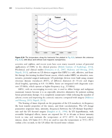
284   Computational Modeling in Biomedical Engineering and Medical Physics


                Figure 8.26 The temperature along the horizontal line plotted in Fig. 8.25, between the antennae
                (Fig. 8.25), with (blue) and without (red) magnetic nanoparticles.
                seventies and eighties, and recent years have seen many research venues of potential
                applications of HIFU in the clinical practice (British Institute of Radiology, 2014).
                Preclinical, and clinical studies (Furusawa et al., 2007; Maloney and Hwang, 2015;
                Napoli, 2013), and tests on US devices suggest that HIFU is a safe, effective, and feasi-
                ble therapy for treating localized breast cancer, which makes HIFU an attractive, non-
                invasive, potential surgical instrument. US prototype devices were built using ceramic
                zirconate titanate transducers (PZT), of different diameters (8 10 cm) and shapes
                (focal lengths), operating at 0.8 1.6 MHz. Some are integrated with diagnostic scan-
                ners 3.5 MHz, which operate at higher frequency (e.g., 3.5 MHz).
                   HIFU, with an encouraging recovery rate, is used to ablate benign and malignant
                mammary tumors because it is an especially attractive alternative for patients seeking
                breast preservation therapy. It is completely noninvasive while reducing the number of
                adverse events and improving the cosmetic outcome compared with surgery (Maloney
                and Hwang, 2015; Napoli, 2013).
                   The heating of tissue depends on the properties of the US transducer, its frequency
                the heat transfer properties of the tissues, and their vascularization. The US dosage
                parameters (exposure time, intensity, frequency) between the US therapy hyperther-
                mia and the US focused surgery (Sanghvi et al., 1996). In US hyperthermia, with
                reversible biological effects, tissues are exposed for 10 to 60 min at lower intensity
                levels to raise and maintain the temperature at 41 C 45 C. In focused surgery


                intense, short, US bursts (0.1 30 s) are used to raise the temperature at 70 C 90 C


                within a few seconds, to the US ablate the focused tissue volume.