Page 296 - Computational Modeling in Biomedical Engineering and Medical Physics
P. 296
Hyperthermia and ablation  285


                   The ultrasound work interactions

                   US heating aims to reach a temperature of 42 C 45 C in the tumor tissue,

                   which is large enough to kill tumor cells while minimizing the damage produced
                   to the surrounding tissues (Ter Harr, 2007; Solovchuk et al., 2012). For har-
                   monic working conditions, the US propagation mode may be represented in the
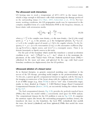
                   complex simplified form of a scalar Helmholtz PDE in the frequency domain, in
                   linear elastic with attenuation media

                                                              2
                                               1             k p t
                                                              eq
                                           r     ð rp t 2 qÞ 2   5 Q;                    ð8:23Þ
                                              ρ               ρ
                                                c              c
                               2
                   where ρ 5  ρc 2 is the complex mass density, ρ is the mass density, c [m/s] is the sound
                          c   c c
                                                                                             2
                                                                                    2
                   speed, p t 5 p 1 p b , is the pressure, p b is the background pressure, k 5 ðω=c c Þ ,
                                                                                    eq
                   c c 5 ω/k is the complex speed of sound, ω 5 2πf is the angular velocity, f is the fre-
                   quency, k 5 ω/c jα is the wavenumber [1/m], α is the attenuation coefficient [Np/
                              3                            -2
                   m], q [N/m ] is a dipole source, and Q [s ] is a monopole source. There is no
                   domain (monopole or dipole) sources in this study.
                      For the part of the boundary which model the transducer a normal acceleration
                                          1                     2
                   condition is set, 2nUð2 ð rp t ÞÞ 5 a n , a n 52 d 0 ω , where d 0 is the maximum dis-
                                         ρ c
                   placement, of the order O(nm)—here 4.56 nm. Perfectly matched layers (PML)—
                   cylindrical for the water and torso, and spherical for the cup—with hard sound
                   boundary conditions (no displacement) close the US problem.

                   Ultrasound ablation of a breast tumor
                   As in thermal therapies, in general, numerical modeling may contribute to the
                   success of the US therapy, providing useful insights in the preinterventional stage.
                   To this aim, a patient-specific computational domain is required, and the first step is
                   the imaging reconstruction of the breast along with the malignant tumor (3DSlicer).
                   A set of MRI images (e.g., from TCIA)isusedas “raw” data. Construction stages,
                   similar to those detailed in Chapter 3: CAD/Medical Image Based Constructed
                   Computational Domains (Baerov, 2019), aresuccessivelyleadingthevolumeshown
                   in Fig. 8.27.
                      The final computational domain, Fig. 8.28, provides the perfectly matched layers
                   (PMLs) that close the model within a conveniently sized space for FEM analysis.
                   The computational domain for the US problem is seen in Fig. 8.28.It comprises
                   the anatomic region (the breast), the surrounding propagation medium (water), the
                   transducer (its trace on the boundary), the back PML (cylindrical) that models
                   the torso, the lateral (cylindrical) and front (spherical) PMLs that model the water
                   reservoir.

