
Page 298 - Computational Modeling in Biomedical Engineering and Medical Physics
P. 298
Hyperthermia and ablation 287
Table 8.6 Material properties used in the US analysis.
Medium Density, Sound Attenuation, Frequency Specific Thermal
3
ρ (kg/m ) speed, c α (Np/m MHz) dependence heat conductivity, k
(m/s) of α (J/kg K) (W/m K)
Water 1000 1522 0.025 f 2 4180 0.59
Tissue 1044 1568 8.550 f 1.5 3710 0.56
Tumor 1044 1568 8.550 f 1.5 4070 0.84
US, Ultrasound sources.
The working frequency is 1 MHz. The water pool is a thermostat, maintaining the
breast surface temperature at 37 C. Other properties used in the US and heat transfer
analyses are listed in Table 8.6 (D’Astous et al., 1986; Ter Harr, 2007; Duck, 1990;
wiki; Jin et al., 2014; Preda, 2019). The perfusion rate is taken 6.4 3 10 23 l/s.
The US problem is solved first, and the heat transfer is integrated next. Some not
trivial elements of numerical modeling, for example, the resolution that the FEM
meshes for US and heat transfer models have to have, the type and order of the inter-
polating polynomials that are selected, and the accuracy test that is required for grid-
independent numerical solutions.
The acoustic pressure field inside the breast, as seen through orthogonal slices,
Fig. 8.29, indicates the US propagation pattern. The US field is focused, with a maximum
concentration that is noticeable, perhaps, too deep inside the breast. This region of highest
US focalization is subject to work interaction that may result in heating the frontal part.
The tumor and some surrounding tissue here are prone to hyperthermia and eventually,
ablation provided the therapeutic protocol is set adequately.
Turning to the heat transfer part, once the acoustic intensity is known, the US
heating is solved, and Figs. 8.30 and 8.31 unveil the temperature inside the breast after
1800 s of exposure to the US.
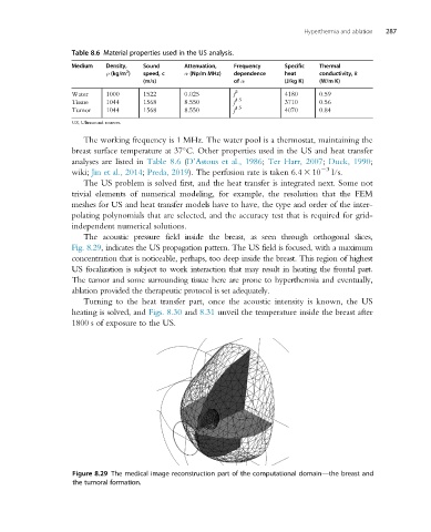
Figure 8.29 The medical image reconstruction part of the computational domain—the breast and
the tumoral formation.