
Page 151 - Computational Retinal Image Analysis
P. 151
2 Automated image quality assessment algorithms 145
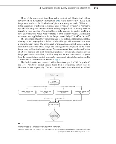
Three of the assessment algorithms (color, contrast and illumination) utilized
the approach of histogram backprojection [51], which assessed how pixels in an
image were similar to the distribution of pixels in a histogram model. With respect
to the assessment of color, for each image class of “bright” or “dark” or “normal” a
specific colourmap was constructed from training images. Each colourmap was used
to perform color indexing of the retinal image to be assessed for quality, resulting in
three color measures which were combined to form a feature vector. Classification
techniques were applied to determine the image class of “bright”, “dark” or “normal”.
The assessment of contrast was also related to the indexing approach and applied
a histogram backpropagation to derive an indexed image, which was used to achieve
a contrast quality score. The assessment of illumination assessed homogeneity in
illumination across the retinal image and a histogram backprojection of the retinal
image using an illumination colourmap. The assessment of focus used a combination
of a Sobel operator and multi-focus-level analysis. The final classification into an
image quality assessment binary decision integrated the previous measures computed
from the image that represented image color, focus, contrast and illumination quality.
An overview of the method can be seen in Fig. 2.
The final classifier was evaluated with a dataset composed of 848 “ungradable”
and 1184 “gradable” retinal images taken from a proprietary dataset and the
Messidor dataset respectively. The best overall results were obtained by a Feed-
Retinal Image
Image
Pre-Processing
Pre-processed Retinal Image Pre-processed Retinal Image
Colour Assessment Focus Assessment Contrast Assessment Illumination Assessment
Algorithm Algorithm Algorithm Algorithm
Focus Measures Contrast Measures
Colour Measures Illumination Measures
Quality Classifier
Overall Retinal
Image Quality
FIG. 2
Retinal image quality assessment algorithm flowchart.
From J.M. Pires Dias, C.M. Oliveira, L.A. da Silva Cruz, Retinal image quality assessment using generic image
quality indicators, Inf. Fusion 19 (2014) 73–90.