
Page 306 - Computational Retinal Image Analysis
P. 306
304 CHAPTER 15 Retinal biomarkers and cardiovascular disease
(A)
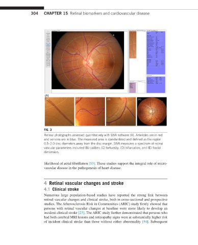
FIG. 3
Retinal photographs assessed quantitatively with SIVA software (A). Arterioles are in red
and venules are in blue. The measured area is standardized and defined as the region
0.5–2.0 disc diameters away from the disc margin. SIVA measures a spectrum of retinal
vascular parameters included (B) caliber; (C) tortuosity; (D) bifurcation; and (E) fractal
dimension.
likelihood of atrial fibrillation [53]. These studies support the integral role of micro-
vascular disease in the pathogenesis of heart disease.
4 Retinal vascular changes and stroke
4.1 Clinical stroke
Numerous large population-based studies have reported the strong link between
retinal vascular changes and clinical stroke, both in cross-sectional and prospective
studies. The Atherosclerosis Risk in Communities (ARIC) study firstly showed that
persons with retinal vascular changes at baseline were more likely to develop an
incident clinical stroke [23]. The ARIC study further demonstrated that persons who
had both cerebral MRI lesions and retinopathy signs were at substantially higher risk
of incident clinical stroke than those without either abnormality [54]. Subsequent