
Page 305 - Computational Retinal Image Analysis
P. 305
3 Retinal vascular changes and heart disease 303
(A) (B)
(C)
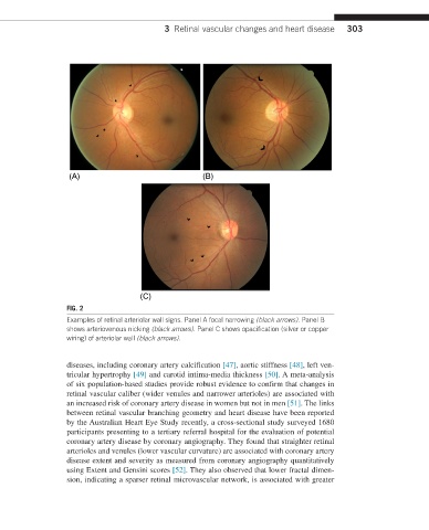
FIG. 2
Examples of retinal arteriolar wall signs. Panel A focal narrowing (black arrows). Panel B
shows arteriovenous nicking (black arrows). Panel C shows opacification (silver or copper
wiring) of arteriolar wall (black arrows).
diseases, including coronary artery calcification [47], aortic stiffness [48], left ven-
tricular hypertrophy [49] and carotid intima-media thickness [50]. A meta-analysis
of six population-based studies provide robust evidence to confirm that changes in
retinal vascular caliber (wider venules and narrower arterioles) are associated with
an increased risk of coronary artery disease in women but not in men [51]. The links
between retinal vascular branching geometry and heart disease have been reported
by the Australian Heart Eye Study recently, a cross-sectional study surveyed 1680
participants presenting to a tertiary referral hospital for the evaluation of potential
coronary artery disease by coronary angiography. They found that straighter retinal
arterioles and venules (lower vascular curvature) are associated with coronary artery
disease extent and severity as measured from coronary angiography quantitatively
using Extent and Gensini scores [52]. They also observed that lower fractal dimen-
sion, indicating a sparser retinal microvascular network, is associated with greater