
Page 303 - Computational Retinal Image Analysis
P. 303
2 The concept of retinal vascular imaging 301
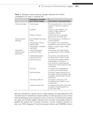
Panel 1 Glossary of retinal vascular changes measured from retinal
photographs and used in studying CVD.
Parameters measured
from retinal imaging Descriptions of the parameters
Retinopathy signs Hemorrhages Red deposits with dot, blot or flame
shaped caused by exudation of
blood
Exudates White or yellow deposits with
distinct margin caused by
exudation of lipids
Cotton-wool spots Fluffy white lesions caused by
ischemia of nerve fiber layer
Retinal arteriolar Focal arteriolar narrowing Focal narrowing or constriction of
wall signs (FAN) retinal arterioles
Arteriovenous nicking/ Compression of the venules by
nipping (AVN) arterioles at arteriovenous crossing
Opacification of arteriolar Silver or copper wiring of retinal
wall (OAW) arterioles
Quantitative Central retinal artery A summary index reflecting the
retinal vascular equivalent (CRAE) average width of retinal arterioles
parameters
Central retinal venule A summary index reflecting the
equivalent (CRVE) average width of retinal venules
Fractal dimension A measure of a fractal structure,
that exhibits the property of
self-similarity, characterizing the
distribution of the branching retinal
vasculature
Tortuosity A measure of the straightness/
curliness of the retinal vessels
Branching angle An angle measure defined as the
first angle subtended between two
daughter vessels at each vascular
bifurcation
Branching coefficient A measure of the ratio of the
branching vessel widths to trunk
vessel width
Length-diameter-ratio A measure of the ratio of the length
between 2 branching points to
trunk vessel width
efficiency and thereby, a greater risk of vascular damage, providing additional CVD
risk information to enable better predictive ability of cardiovascular outcomes. Newer
retinal branching geometric parameters such as tortuosity, fractal dimension, branch-
ing angles and vascular length-to-diameter ratio were thus subsequently defined to
quantify the optimal state of retinal vasculature in recent computer software sys-
tems (e.g., SIVA (Singapore I Vessel Assessment) software and VAMPIRE software