
Page 304 - Computational Retinal Image Analysis
P. 304
302 CHAPTER 15 Retinal biomarkers and cardiovascular disease
(A) (B)
(C) (D)
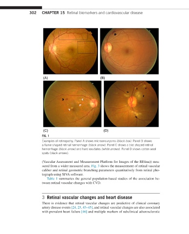
FIG. 1
Examples of retinopathy. Panel A shows microaneurysms (black box). Panel B shows
a flame shaped retinal hemorrhage (black arrow). Panel C shows a blot shaped retinal
hemorrhage (black arrow) and hard exudates (white arrows). Panel D shows cotton wool
spots (black arrows).
(Vascular Assessment and Measurement Platform for Images of the REtina)) mea-
sured from a wider measured area. Fig. 3 shows the measurement of retinal vascular
caliber and retinal geometric branching parameters quantitatively from retinal pho-
tograph using SIVA software.
Table 1 summaries the general population-based studies of the association be-
tween retinal vascular changes with CVD.
3 Retinal vascular changes and heart disease
There is evidence that retinal vascular changes are predictive of clinical coronary
artery disease events [24, 25, 43–45], and retinal vascular changes are also associated
with prevalent heart failure [46] and multiple markers of subclinical atherosclerotic