
Page 72 - Computational Retinal Image Analysis
P. 72
4 Retinal image registration 63
eye motion) complicate image registration further, due to projective distortion of the
curved surface of the eye.
Applications of RIR can be classified according to whether images are acquired
in the same or different examinations. Images from the same examination are devoid
of anatomic changes. If their overlap is significant, they can be combined into images
of higher resolution and definition [88–90], enabling more accurate measurements.
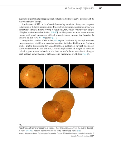
Images with small overlap are utilized to create image mosaics that broaden the
sensor’s field of view [91–93] (see Fig. 1).
Longitudinal studies of the retina [97, 98] are facilitated by the registration of
images acquired at different examinations (i.e., initial and follow-up). Pertinent
studies enable disease monitoring and treatment evaluation, through tracking of
symptom reversal. In this context, accurate registration of images of the same
retinal region proves valuable in the detection of minute but critical changes,
such as local hemorrhages or differences in vasculature width (see Fig. 2).
FIG. 1
Registration of retinal images into a mosaic. Top: Original images, from the public dataset
in Refs. [94, 95]. Bottom: Registration result, using Hernandez-Matas [96].
From C. Hernandez-Matas, Retinal Image Registration Through 3D Eye Modelling and Pose Estimation (Ph.D.
thesis), University of Crete, 2017.